On the theory and practice of midwifery / by Fleetwood Churchill.
- Fleetwood Churchill
- Date:
- 1866
Licence: Public Domain Mark
Credit: On the theory and practice of midwifery / by Fleetwood Churchill. Source: Wellcome Collection.
16/854 page 4
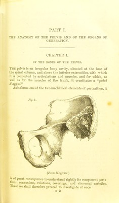
![In the adult, tlie pL4vis may bo divided into four parts or bones; viz., two ossa innominala, the o„- sacrum, and the os coccygis; but in early life they are more minutely divisible. Let us now examine these bones separately. Each Os Innojiinatitm, at an early period of intra-uterine life, consists of cartilage only, in which, subscrpicnfly, numerous spicula; of ossification are seen, and which at birth have coalesced so as to ibrm three bones, separated by cartilage. After birth, the process of ossification continues until these separate bones meet in the acetabulum, where they are identified with each other, and at the symphysis pubis, where the opposite ossa pubis are united by cartilage and ligaments. The breadth of each os innominatnm, from the anterior-superior to the posterior- superior spinous process, is six inches, and the height from the tuber ischii to the highest jjart of the crest of the ilium is seven inches. The three bones into which each os innominatnm is divided at birth have received difterent names, and require a distinct notice. The Os Ilium, hip, or hauncli-bone (figs. 1, 2) is the larger of the three, of a triangular shape, situated superiorly, and with its fellow forming what is called the false pelvis. Its external surface ('), or dorsum, is convex, ii-regular, with elevations and depressions which serve for the attachment of the glutrei muscles. Its internal surface, or venter ('), is concave and smooth, aflx)rding a bed for the iliacus internus muscle. The lotver portion, body or base (■'), is the thickest part of the bone, and forms more than one-third of the acetabulum. Above the body, the bone spreads out into its ala or wing, which rises obliquely forwards, upwards, outwards, and then backwards, ter- minating in the crest or cQ-ista ilii—a semicircular ridge of some thickness, which, at its posterior part, curves downwards and forwards. Its borders serve for the attachment of the abdo- minal muscles, and certain ligaments to be hereafter described ; and it terminates anteriorly, in the anterior-superior, and an- terior-inferior spinous process (•* The former affords attach- ment to Poupart's ligament, the tensor vaginas femoris, the sar- torius, and a portion of the rectus femoris muscles. Between the posterior spinous processes is a deep arch,—the sciatic notch —which is divided by ligaments into the two sciatic ibraniina: through the upper of these, which is the larger, pass the gluteal, sciatic, and pudic arteries, the sciatic and pudic nerves, and tlio jjyrilorm muscle; whilst, through the inferior opening, the ])udic arteries and nerve re-enter the pelvis, and the obturator internus muscle passes out. The posterior part of the crest of the ilium](https://iiif.wellcomecollection.org/image/b20398864_0016.jp2/full/800%2C/0/default.jpg)


 No text description is available for this image
 No text description is available for this image
 No text description is available for this image