Volume 1
A system of human anatomy : including its medical and surgical relations / by Harrison Allen. With a section on histology. By E.O. Shakespeare.
- Harrison Allen
- Date:
- 1882-1884
Licence: Public Domain Mark
Credit: A system of human anatomy : including its medical and surgical relations / by Harrison Allen. With a section on histology. By E.O. Shakespeare. Source: Wellcome Collection.
Provider: This material has been provided by The University of Leeds Library. The original may be consulted at The University of Leeds Library.
27/110 page 35
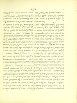
![CONNECTIVE- tlie other liand, tlie vital proclivities of the first men- tioned very thin hyaline cell-plates seem to be entirely dormant. These granular cells are said by Klein and some others to be germinating, and have been called by them germinating endoihelia (see e, fig. 1, Plate I.). They are generally arranged around the mouth of a vertical canal connecting a lympli-channel of the sub jacent connective-tissue with the serous cavity. The activity of this germinating endothelium may be such that the granular cells proliferate and form new-cells, some of which may be set free in the serous cavity. These new cells are similar in every respect to the other lymph-corpuscles which float in the lympli- plasma, and cannot, after separation from their place of generation, be distinguished from them. The endothelia covering some portions of the abdominal cavities and the fenestrated septa of the mediastinum are thus a very fruitful source of supply of the color- less corpuscles of the lym]>h. The endothelia are, as already stated, somewhat elastic. When the membrane which they cover is stretched, they become thinner, and spread out in order to cover a wider extent of surface, and their border becomes straighter. On the contrary, when the membrane retracts, their broad diameter lessens, their borders become more sinuous, and the cell be comes thicker. Thus during full inspiration the endo- thelia covering the pleural surface of the lungs are much thinner and broader than at the end of expira- tion. At the latter instant the pulmonary endothelia are quite cubical in shape. As has already been indicated, the endothelia not only of the serous cavities, and of the lymph-vessels, but also of the bloodvessels as well, may be regarded as some of the sources of the colorless elements to the lymph and blood. CONNECTIVE-TISSUE CELLS. Still another group of the elements derived from the mesoblast is formed by the cells of the connective- tissue. They may be considered under two general heads—the wandering cells and the fixed cells. Wandering cells.—The wandering cells of the connec- tive-tissue are not different from the colorless cells already described when considering the lymph. They are in reality lymph-corpuscles, existing in the radi- cles of the lymphatic system, and do not call for any particular description here. One form may be con- sidered for a moment, however, before dismissing them. There are found in the interstices of the loose connective-tissues which are especially vascular, small TISSUE CELLS. 35 numbers of large granular corpuscles slightly pig- mented (plasmatic corpuscles of Waldeyer). They are more frequent along the walls of vessels, and do not usually exhibit marked amoeboid movements. Connective-tissue corpuscles.—The fixed cells of the connective-tissue vary much in shape according to the arrangement of the fibrous tissue in which they are imbedded. The cellular elements of the great connective-tissue system are met with only within or upon the sides of the spaces which permeate that system, whether they be in the form of fine channels or in the nature of lymph-cavities, and whether those spaces be large or small. The large spaces, as, for example, the perito- neal cavity, the pleural cavity, the arachnoid cavity, the heart and bloodvessels, the larger lymph-vessels, are lined throughout by a complete covering of endo- thelial cells; the minute spaces which exist between the bundles of connective-tissue fibrils have as a rule only a more or less incomplete lining. But whether the space be of one kind or of the other, it is lined by cells of an essentially identical nature; only their forms and the arrangement of some of their structures, vary according to the circumstances which surround them. Although the fixed cells of the connective- tissue are here considered under a special section dif- ferent from that in which the endothelia were dis- cussed, it is not because these two species of cells differ in anything more than form. The so-called fixed cell of the connective tissue tends to assume a form which is a more or less perfect mould of the space in which it is found. It presents two general forms, the one plate-like, the other stel- late. The plate-like forms more nearly resemble the cell-plates of the endothelia than do the stellate forms, and will therefore be considered first (see c, fig. 3, PI. IX.). Flat tendon-cells.—In white fibrous tissue such as tendon, etc., the spaces formed hj the apposition of several bundles are linear, and are limited laterally bv the convex surfaces of the fibrous bundles. The surface of the space thus formed is usually partially lined only. On one side is a longitudinal row of flat elastic cell-plates, applied more or less tightly to the surface of the fibrous bundles forming that side. The opposite side of this minute lymph-space is void of cellular lining. The cells constituting this row are placed edge to edge, and at the line of junction the border of the cell is quite straight, and generally transverse to the direction of the bundle. These cells thus have two long straight parallel sides. The lateral borders of the cell are more or less notched,](https://iiif.wellcomecollection.org/image/b21506607_0001_0027.jp2/full/800%2C/0/default.jpg)



 No text description is available for this image
 No text description is available for this image
 No text description is available for this image