Volume 1
A system of the anatomy of the human body. Illustrated by upwards of two hundred and fifty tables, taken partly from the most celebrated authors, and partly from nature / By Andrew Fyfe.
- Andrew Fyfe
- Date:
- 1820
Licence: Public Domain Mark
Credit: A system of the anatomy of the human body. Illustrated by upwards of two hundred and fifty tables, taken partly from the most celebrated authors, and partly from nature / By Andrew Fyfe. Source: Wellcome Collection.
349/372
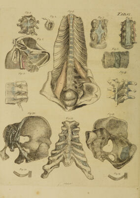
![the first and second Phalanges of the Fingers. They run obliquely forwards within the Vaginal Ligaments, terminate in the Tendons of the Two Flexor Muscles of the Fingers, and assist in keeping them in their places. Tab. LXII. Fig. ] 8.f g, h. The External Transverse, or Posterior Annular Liga¬ ment of the Wrist, which is part of the Aponeurosis of the Fore-arm, extending across the back of the Wrist, from the inner side of the extremity of the Ulna and Os Pisiforme, to the outer side of the extremity of the Ra¬ dius. Tab. XL. Fig. 2. E. It is connected with the small Annular Ligaments which tie down the Tendons of the Extensores Ossis Metacarpi et Primi Internodii Pollicis, and the Extensor Carpi Ulnaris. Tab. LVI-II. Fig. 5. i, l. The Vaginal Ligaments, which adhere to the former Ligaments, and serve as Sheaths and Bursae Mucosae to the Extensor Tendons of the Hand and Fingers. Tab. LIX. Fig. 2. The Transverse Ligaments of the Extensor Tendons, which are Aponeurotic Slips running between the Ten¬ dons of the Extensor Digitorum Communis, near the Heads of the Metacarpal Bones, and retaining the Ten¬ dons in their places. Tab. LVIII. Fig. 5. o. LIGAMENTS OF THE INFERIOR EXTREMITIES. Ligaments, &c. connecting the Os Femoris with the Os Innominatum. The Capsular Ligament, the largest and strongest of the Articular Ligaments. It arises round the outside of the Brim of the Acetabulum, embraces the Head of the Thigh-bone, and incloses the whole of its Cervix as far as the root or outer extremity, round which it is firmly connected. Tab. XXXVIII. Fig. 18,'E. The outer part of the Capsular Ligament is extended farther down than the inner, which is reflected back upon the Neck of the Bone, and in certain parts forms Retinacula. Tab. LXIII. Fig. 1. 2. It is not every where of the same strength. It is thickest at its anterior and outer part; thinner where it is covered by the Iliacus Internus; and thinnest poste¬ riorly) where the adjacent Quadratus is opposed to it. It is strengthened on its outer Surface by various Ac¬ cessory or Additional Slips, which run down from the Fascia Lata and surrounding Muscles; but the strongest of these Slips arises with diverging Fibres from the in¬ ferior-anterior Spinous Process of the Os Ilium. Tab. LXIII. Fig. 1. m, n, o. The Capsular Ligament allows the Thigh-bone to be moved to every side; and when its Body is brought for¬ wards or backwards, a small degree of rotation is per¬ formed by the Cervix of the Bone, round its own axis. The Internal, commonly called the Round Ligament, which arises by a broad flat beginning from the under and inner part of the Cavity ot the Acetabulum, and is connected with the Substance termed Gland of the Joint. From this it runs backwards and a little upwards, be¬ coming gradually narrower and rounder, to be fixed to the Pit upon the inner Surface of the Ball of the Os Femoris. 'Fab. LXIII. Fig. 2. g—Jc. The Round Ligament prevents the Bone from bein dislocated upwards or inwards, and assists in agitatin the Mucous Substance within the Joint. VOL. I. A Cartilaginous Ligament surrounding the Brim of the Acetabulum, and thereby increasing the depth of the Cavity for the reception of the Head of the Thigh¬ bone. Tab. LXIII. Fig. 2. c. A Double Cartilaginous Ligament, Tab. LXIII. Fig. 3. d, stretched from one end of the Breach in the un¬ der and fore part of the Acetabulum to the other, but leaving a Hole behind it for containing part of the Sub¬ stance called Gland of the Joint, and for the passage of the Vessels of that Substance. This Ligament allows the Thigh-bone to be moved inwards, and the Glandular-looking Substance to be agitated with safety. The Substance called Gland of the Joint, covered with a Vascular Membrane, and lodged in a Depression in the under and inner part of the Acetabulum. Tab. LXIII. Fig. 2. m. At the edges of this Substance Fringes are sent out, which furnish part of the Synovia for the lubrication of the Joint. The edges of this Substance are fixed to those of the Pit in the Acetabulum, by small Ligamentous Bridles, termed Ligamenta Mucosa, vel Ligamentula Massce A- diposo-glandulosce. Ligaments, &c. of the Joint of the Knee. The Lateral Ligaments which lie at the sides of the Joint, and adhere to the outer Surface of the Capsular Ligament. The Internal Lateral Ligament, which is of consi¬ derable breadth, arising from the upper part and Tu¬ bercle of the Internal Condyle of the Os Femoris, and inserted into the upper and inner part of the Tibia; the Fibres passing obliquely forwards, till they have reached a little below the Head of the Bone. Tab. LXIII. Fig. 4. Jc.](https://iiif.wellcomecollection.org/image/b30455443_0001_0349.jp2/full/800%2C/0/default.jpg)