The anatomy of the nervous system : from the standpoint of development and function / by Stephen Walter Ranson ... with 284 illustrations, some of them in colors.
- Stephen Walter Ranson
- Date:
- 1927
Licence: In copyright
Credit: The anatomy of the nervous system : from the standpoint of development and function / by Stephen Walter Ranson ... with 284 illustrations, some of them in colors. Source: Wellcome Collection.
34/432 (page 34)
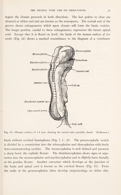
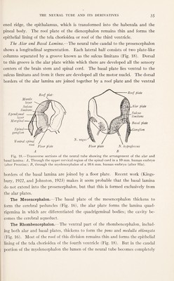
![lamina is a portion of the telencephalic cavity which forms the anterior part of the third ventricle. The further development of these structures is readily traced in Fig. 16, which represents the brain of a human fetus of the third month. The most striking feature of the brain at this stage is the great size attained by the cerebral hemispheres. The Diencephalon.—The three principal divisions of the diencephalon— the thalamus, epithalamus, and hypothalamus—faintly indicated in an embryo of 13.6 mm., are well defined by the third month (Fig. 16). In transverse sec¬ tions this division of the embryonic brain is seen to be composed of a pair of plates on either side, which with a roof and floor form the walls of the ventricle (Fig. 17). The more dorsal members of each pair of lateral plates become greatly thickened and form the thalamus, while the more ventral ones form the hypo¬ thalamus. On either side these plates meet at an angle, forming the hypothalamic sulcus. Table Showing Subdivisions of the Neural Tube and Their Derivatives (Modified from a Table in Keibel and Mall, Human Embryology). Primary vesicles. Subdivisions. Derivatives. Lumen. / j / ! Telencephalon. Cerebral cortex, Corpora striata, Rhinencephalon, Pars-optica hypo¬ thalami. Lateral ventricles. Rostral portion of the third ventricle. Brain.< Prosencephalon... . < V Diencephalon. Epithalamus, Thalamus, Hypothalamus, Hypophysis, Tuber cinereum, Mammillary bodies, Metathalamus. The greater part of the third ventricle. Mesencephalon ( Mesencephalon. . .. < Corpora quadri- gemina, Crura cerebri. Cerebral aqueduct. \ / Rhombencephalon j Metencephalon j Myelencephalon Cerebellum, Pons, Medulla oblongata.] Fourth ventricle. Spinal cord Spinal cord. Central canal. The hypothalamus gives rise to the tuber cinereum, posterior lobe of the hy¬ pophysis, and the mammillary bodies. From the dorsal edge of the thalamic lamina, where this is attached to the thin roof plate, there is developed a thick-](https://iiif.wellcomecollection.org/image/b29813670_0034.jp2/full/800%2C/0/default.jpg)