Anatomy, descriptive and surgical / by Henry Gray ; the drawings by H.V. Carter ; the dissections jointly by the author and Dr.Carter.
- Henry Gray
- Date:
- 1860
Licence: Public Domain Mark
Credit: Anatomy, descriptive and surgical / by Henry Gray ; the drawings by H.V. Carter ; the dissections jointly by the author and Dr.Carter. Source: Wellcome Collection.
31/838
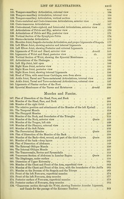
![FI<3. PAGE 121. Temporo-maxillary Articulation, external view 164 122. Temporo-maxillary Articulation, internal view 164 123. Temporo-maxillary Articulation, vertical section 165 124. Costo-vertebral and Costo-transverse Articulations, anterior view . . . 166 125. Costo-transverse Articulations Arnold 167 126. Costo-sternal, Costo-xiptioid, and Intercostal Articulations, anterior view . 169 127. Articulations of Pelvis and Hip, anterior view 170 128. Articulations of Pelvis and Hip, posterior view .171 129. Vertical Section of the Symphysis Pubis 174 130. Sterno-clavicular Articulation 175 131. Shoulder Joint, Scapulo-clavicular Articulation, and proper Ligaments of Scapula 177 132. Left Elbow Joint, showing anterior and internal Ligaments . . . .180 133. Left Elbow Joint, shewing Posterior and external Ligaments .... 181 134. Ligaments of Wrist and Hand, anterior view .... Aiiiold 183 135. Ligaments of Wrist and Hand, posterior view .... do. 183 136. Vertical Section of Wrist, shewing the Synovial Membranes .... 187 137. Articulations of the Phalanges ] 88 138. Left Hip Joint, laid open . . . . , 189 139. Right Knee Joint, anterior view 191 140. Right Knee Joint, posterior view 191 141. Right Knee Joint, shewing internal Ligaments 193 142. Head of Tibia, with semi-lunar Cartilages, seen from above .... 193 143. Ankle Joint, Tarsal and Tarso-metatarsal Articulations, internal view . . 196 144. Ankle Joint, Tarsal and Tarso-metatarsal Articulations, external view . . 197 145. Ligaments of Plantar surface of the Foot 199 146. Synovial Membranes of the Tarsus and Metatarsus . . . Arnold 200 Muscles and Fascise. 147. Plan of Dissection of the Head, Face, and Neck 207 148. Muscles of the Head, Face, and Neck 208 149. Muscles of the right Orbit 212 150. The relative position and attachment of the Muscles of the left Eyeball . . 212 151. The Temporal Muscle 218 152. The Pterygoid Muscles 219 153. Muscles of the Neck, and Boundaries of the Triangles 223 154. Muscles of the Neck, anterior view Quain 225 155. Muscles of the Tongue, left side . 229 156. Muscles of the Pharynx, external view 230 157. Muscles of the Soft Palate 232 158. The Prevertebral Muscles Quain 235 159. Plan of Dissection of the Muscles of the Back 238 160. Muscles of the Back—first, second, and part of the third layers Quain 239 161. Muscles of the back—deep layers 243 162. Plan of Dissection of Abdomen 249 163. The External Oblique Muscle 250 164. The Internal Oblique Muscle 251 165. The Transversalis, Rectus and Pyramidalis 253 166. Transverse Section of Abdomen in Lumbar Region . . . Quain 254 167. The Diaphragm, under surface 258 168. Dissection of Upper Extremity , 262 169. Muscles of the Chest and Front of the Arm, superficial view .... 263 170. Muscles of the Chest and Front of the Arm, with the boundaries of the Axilla 265 171. Muscles on the Dorsum of the Scapula and the Triceps 269 172. Front of the left Fore-arm, superficial muscles 274 173. Front of the left Fore-arm, deep muscles 277 174. Posterior surface of Fore-arm, superficial muscles 279 175. Posterior surface of Fore-arm, deep muscles 282 176. * Transverse section through the Wrist, shewing Posterior Annular Ligament, and Canals for the passage of the Extensor Tendons 283](https://iiif.wellcomecollection.org/image/b20386424_0031.jp2/full/800%2C/0/default.jpg)


 No text description is available for this image
 No text description is available for this image
 No text description is available for this image