Anatomy, descriptive and surgical / by Henry Gray ; the drawings by H.V. Carter ; the dissections jointly by the author and Dr.Carter.
- Henry Gray
- Date:
- 1860
Licence: Public Domain Mark
Credit: Anatomy, descriptive and surgical / by Henry Gray ; the drawings by H.V. Carter ; the dissections jointly by the author and Dr.Carter. Source: Wellcome Collection.
76/838 page 40
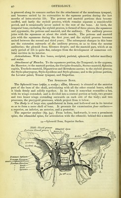
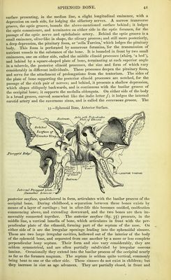
![is grooved along its concave surface for the attachment of the membrana tympani, and becomes united by its extremities to the sqamous portion during the last months of intra-uterine life. The petrous and mastoid portions then become ossified, and lastly the styloid process, which remains separate a considerable period, and is occasionally never united to the rest of the bone. At birth, the temporal bone, excluding the styloid process, is formed of three pieces, the squamous and zygomatic, the petrous and mastoid, and the auditory. The auditory process joins with the squamous at about the ninth month. The petrous and mastoid join with the squamous during the first year, and the styloid process becomes united between the second and third years. The subsequent changes in this bone are the extension outwards of the auditory process, so as to form the meatus auditorius; the glenoid fossa becomes deeper; and the mastoid part, which at an early period of life is quite flat, enlarges from the development of numerous cel- lular cavities in its interior. Articulations. With five bones, occipital, parietal, sphenoid, inferior maxillary and malar. Attachment of Muscles. To the squamous portion, the Temporal; to the zygoma, the Masseter; to the mastoid portion, the Occipito-frontalis, Sterno-mastoid, Splenius capitis, Trachelo-mastoid, Digastricus and Retrahens aurem; to the styloid process, the Stylo-pharyngeus, Stylo-hyoideus and Stylo-glossus; and to the petrous portion, the Levator palati. Tensor tympani, and Stapedius. The Sphenoid Bone. The Sphenoid bone {(T(pr]v, a wedge; eiBo<;^ likeness) is situated at the anterior part of the base of the skull, articulating with all the other cranial bones, which it binds firmly and solidly together. In its form it somewhat resembles a bat, with its wings extended; and is divided into a central portion or body, two greater and two lesser wings extending outwards on each side of the body; and two processes, the pterygoid processes, which project from it below. The Body is of large size, quadrilateral in form, and hollowed out in its interior so as to form a mere shell of bone. It presents for examination four surfaces— a superior, an inferior, an anterior, and a posterior. The superior surface (fig. 34). From before, backwards, is seen a prominent spine, the ethmoidal spine, for articulation with the ethmoid; behind this a smooth](https://iiif.wellcomecollection.org/image/b20386424_0076.jp2/full/800%2C/0/default.jpg)
 No text description is available for this image
 No text description is available for this image
 No text description is available for this image