Paralyses : cerebral bulbar and spinal: a manual of diagnosis for students and practitioners.
- Henry Charlton Bastian
- Date:
- 1886
Licence: Public Domain Mark
Credit: Paralyses : cerebral bulbar and spinal: a manual of diagnosis for students and practitioners. Source: Wellcome Collection.
Provider: This material has been provided by UCL Library Services. The original may be consulted at UCL (University College London)
35/732 page 15
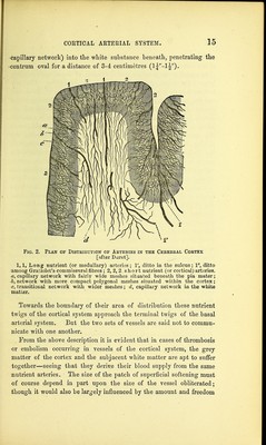
![capillary network) into the white substance beneath, penetrating the centrum oval for a distance of 3-4 centimetres (1J-!^)* Fig. 2. Plan of Distribution of Arteries in the Cerebral Cortex [after Duret]. 1,1, Long nutrient (or medullary) arteries; V, ditto in the sulcus; 1, ditto amoug Gratiolet's commissural fibres ; 2, 2, 2, short nutrient (or cortical) arteries^ a, capillary network with fairly wide meshes situated beDeath the pia mater; b, network with more compact polygonal meshes situated within the cortex; c, transitional network with wider meshes; d, capillary network in the white matter. Towards the boundary of their area of distribution these nutrient twigs of the cortical system approach the terminal twigs of the basal arterial system. But the two sets of vessels are said not to commu- nicate with one another. From the above description it is evident that in cases of thrombosis •or embolism occurring in vessels of the cortical system, the grey matter of the cortex and the subjacent white matter are apt to suffer together—seeing that they derive their blood supply from the same nutrient arteries. The size of the patch of superficial softening must of course depend in part upon the size of the vessel obliterated; though it would also be largely influenced by the amount and freedom](https://iiif.wellcomecollection.org/image/b21270478_0035.jp2/full/800%2C/0/default.jpg)