Paralyses : cerebral bulbar and spinal: a manual of diagnosis for students and practitioners.
- Henry Charlton Bastian
- Date:
- 1886
Licence: Public Domain Mark
Credit: Paralyses : cerebral bulbar and spinal: a manual of diagnosis for students and practitioners. Source: Wellcome Collection.
Provider: This material has been provided by UCL Library Services. The original may be consulted at UCL (University College London)
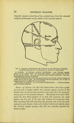
38/732 page 18
![along the superior curved line of the occipital bone, from the external occipital protuberance to the middle of the external meatus. Fig. 3. Diagram illustrating the position of the Principal Cerebral Fissures relative to the Surface of the Scalp [after Reid]. A, Glabella. B, External occipital protuberance, eap, External angular process of the frontal bone. BC, Transverse fissure. AB, Longitudinal fissure. Sy. fis., Sylvian fissure. Sy. h. fis., Horizontal limb of fissure of Sylvius. Sy. a. fis., Ascending limb of fissure of Sylvius. DE, Perpendicular line from depression in front of external auditory meatus to middle line of top of head. FGr, Perpendicular line from posterior end of base of mastoid process to middle line of top of head. FH, Fissure of Rolando, p. o. fis., Parieto-occipital fissure. + Most prominent part of parietal eminence. Fissure of Sylvius.—To find this fissure draw a line from a point one inch and a quarter behind the external angular process of the frontal bone, to a point three-quarters of an inch below the most pro- minent part of the parietal eminence. Measuring from before back- wards the first three-quarters of an inch will represent the main fissure, and the rest of the line will indicate the horizontal limb. The ascending limb will start from the posterior end of the line indi- cating the main fissure—that is, two inches behind and slightly above the external angular process, and will run vertically upwards for about an inch.](https://iiif.wellcomecollection.org/image/b21270478_0038.jp2/full/800%2C/0/default.jpg)