Paralyses : cerebral bulbar and spinal: a manual of diagnosis for students and practitioners.
- Henry Charlton Bastian
- Date:
- 1886
Licence: Public Domain Mark
Credit: Paralyses : cerebral bulbar and spinal: a manual of diagnosis for students and practitioners. Source: Wellcome Collection.
Provider: This material has been provided by UCL Library Services. The original may be consulted at UCL (University College London)
650/732 page 626
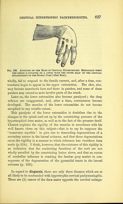
![Still, it is most common for the paralysis to predominate in the muscles supplied by the ulnar and median nerves, and for the atrophy also to be most marked therein. The muscles supplied by the musculo-spiral nerve are decidedly less affected. This, at least, is what happens in the majority of cases, that is, when the lower half of the cervical enlargement is the region most distinctly involved in the morbid process. The result of the above-mentioned predomi- nance of paralysis in certain groups of muscles is the production of a peculiar position of the hand (Fig. 135), in which the hand and the thumb are strongly extended while the fingers are flexed in claw-like fashion. Fig. 135. Attitude of the Hand in Cervical Hypertrophic Pachymeningitis when the disease is on a level with the lower half of the cervical enlargement of the Spinal Cord [after Charcot]. When the stress of the disease occurs, however, at a higher level, that is, opposite the upper half of the cervical enlargement, then it is that the roots of the musculo-spiral nerve, or the corresponding areas of grey matter, become more involved than those of the ulnar and median nerves, and the maximum amount of paralysis also occurs in muscles supplied by the former nerve. The result, as was seen in a remarkable case under the care of Dr. Leech, was to produce an altogether different attitude of the hand and arm. Speaking of this case, Boss says:— The arm is held close to the side, the forearm is extended on the arm and strongly pronated, the hand is flexed on the forearm, the fingers are on a line with or only slightly extended on the metacarpal bones, and the phalanges are extended upon one another, while the thumb is flexed into the palm. (Fig. 136.) The paralysed and atrophied muscles either wholly, or almost](https://iiif.wellcomecollection.org/image/b21270478_0650.jp2/full/800%2C/0/default.jpg)


 No text description is available for this image
 No text description is available for this image
 No text description is available for this image