An atlas of the medulla and midbrain / Edited by H.McE.K.
- Florence R. Sabin
- Date:
- 1901
Licence: In copyright
Credit: An atlas of the medulla and midbrain / Edited by H.McE.K. Source: Wellcome Collection.
Provider: This material has been provided by UCL Library Services. The original may be consulted at UCL (University College London)
15/154 (page 935)
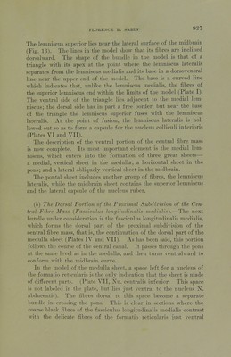
![The middle portion can be related as follows: while the ventral fibres jnake a floor, as it were, at the junction of the pars dorsalis with the pars ventralis pontis, the dorsal fibres remain close to the median line, and into the area thus left between these two fibre masses the middle fibres spread out (Plates Y, VI and VII, For- matio reticularis alba). It is noAv necessary to describe the A'arious portions of the prox- imal subdivision of the central fibre mass more in detail. It will be most convenient in this description to follow (a) the ventral portion, (b) the dorsal portion, and (c) the middle portion separately through the pons and midbrain. (a) The Ventral Portion of the Froxiinal Subdivision of the Cen- tral Fibre Mass (Lemniscus lateralis, Lenfiniscus medialis and Lemniscus superior).—The ventral fibres of the proximal subdi- vision of the central fibre mass make (1) the horizontal pontal sheet and (2) the vertical midbrain sheet. In entering the pons the ventral fibres of the medulla sheet curve rapidly dorsahvard, so that they all pass through the cross-bar (Plate V). As the mass of fibres leaves the cross-bar, it spreads out into the horizontal pontal sheet (Plates V and VI). The lateral portion of this hori- zontal sheet consists of a new mass of fibres w^hich does not exist in the medulla sheet, but is added on in passing through the cross-bar. It forms a part of the pontal sheet for a short distance only, inas- much as it inclines rapidly dorsalward in order to reach the nucleus colliculi inferioris in which a large part of it disappears (Plate I). The space shown in the model between the lemniscus lateralis and the pontal sheet is occupied at this stage of development by indif- ferent substance. The pontal sheet is best seen from its dorsomesial aspect (Plate V). The mesial edge is clearly defined. The sheet is com- paratively thick as it emerges from the trapezoid body, but it grows thinner as it approaches the midbrain. The dorsal surface is level within the pons but curves ventralward in approaching the mid- brain, while the ventral surface shows an anteroposterior curve in crossing the pons (Plate I). The fibres of the medial third of the sheet, its thinnest portion, are cut oif abniptly in the model as they are entering the midbrain in order to accommodate the radix 'N. oculoraotorii. In reality they pass toward the nucleus ruber and appear to form a ])art of its capsule (Plate VI).](https://iiif.wellcomecollection.org/image/b21272050_0015.jp2/full/800%2C/0/default.jpg)