Licence: Public Domain Mark
Credit: Elements of histology / by E. Klein. Source: Wellcome Collection.
72/378 page 56
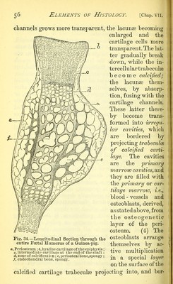
![channels grows more transparent, the lacunae becoming enlarged and the cartilage cells more transparent. The lat- ter gradually break down, while the in- tercellular trabeculse become calcified; the lacunae them- selves, by absorp- tion, fusing with the cartilage channels. These latter there- by become trans- formed into irregu- lar cavities, which are bordered by projecting trabeculce of calcified carti- The cavities are the primary marrow cavities, and they are filled with the primary or car- tilage marrow, i.e., blood - vessels and osteoblasts, derived, as stated above, from the osteogenetic layer of the peri- osteum. (4) The osteoblasts arrange themselves by ac- tive multiplication in a special layer on the surface of the calcified cartilage trabeculse projecting into, and bor- Fig. 34.—Longitudinal Section through the entire Fcetal Humerus of a G-uinea-pig. a, Periosteum ; 6, hyaline cartilage of the epiphysis; - c, intermediate carti]aj?e at the end of the shaft; d, zone of calcification \e, periostealbone,spongy; /, endochondral bone, spongy.](https://iiif.wellcomecollection.org/image/b2041044x_0078.jp2/full/800%2C/0/default.jpg)
 No text description is available for this image
 No text description is available for this image
 No text description is available for this image