Atlas of the external diseases of the eye : including a brief treatise on the pathology and treatment / by O. Haab ; Authorized translation from the German, edited by G.E. de Schweinitz.
- Haab O. (Otto), 1850-1931.
- Date:
- 1899
Licence: Public Domain Mark
Credit: Atlas of the external diseases of the eye : including a brief treatise on the pathology and treatment / by O. Haab ; Authorized translation from the German, edited by G.E. de Schweinitz. Source: Wellcome Collection.
Provider: This material has been provided by the Royal College of Physicians of Edinburgh. The original may be consulted at the Royal College of Physicians of Edinburgh.
154/312 (page 126)
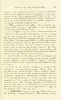
![On the other hand, the deformities of the lids which often resnlt must be corrected by surgical means. Pannns usually requires no special treatment. If the vascnlar tissue is nnusually thick, cauterization may be practised with great care. If ulcers develop in the cornea, co])per snlphate must be used instead of silver nitrate [and the treatment suitable for corneal ulcer instituted.—Ed.]. The patient and his attendants should be duly impressed with the im])ortance of observing proper precautions against the spread of the disease. If possible the patient sliould be isolated, especially if the disease appears in large bodies of men, as in an army. 6. Spring=conjunctivitis (Fruehjahr’s Catarrh). Spring-catarrh is the only process in the human body, with the exception of freckles, that is exclusively depend- ent on atmospheric heat, so much so that it does not attain its full development in cool seasons. It is a diffuse in- flammation, involving the entire conjunctiva, although localized deposits are sometimes observed. The disease is quite rare in some localities, and occurs most frequently in young men, giving them a strikingly pale and languid ajq)earance and often lasting for years. Owing to the slight degree of ptosis which is usually present the patients have the same dull, sleepy look that is seen in trachoma. One characteristic symptom is a peculiar yellowish-red discoloration of the conjunctiva on either side of the cornea (Plate 15, d). The remaining objective j)henomena may be divided into three grouj)s : 1. Hypertrophy at the sclcrocorneal junction, consisting of smooth, semitransparent nodules, of pinkish color and waxy appearance, found chiefly on either side of the cornea, but occasionally encroaching on the upper and lower segments of the limbus (Plate 15, a and d). These nodules never undergo degeneration. 2. The so-called tessellated or pavement-granulations on the tarsal conjunctiva (so called on account of their resem-](https://iiif.wellcomecollection.org/image/b21691587_0154.jp2/full/800%2C/0/default.jpg)