Atlas of the external diseases of the eye : including a brief treatise on the pathology and treatment / by O. Haab ; Authorized translation from the German, edited by G.E. de Schweinitz.
- Haab O. (Otto), 1850-1931.
- Date:
- 1899
Licence: Public Domain Mark
Credit: Atlas of the external diseases of the eye : including a brief treatise on the pathology and treatment / by O. Haab ; Authorized translation from the German, edited by G.E. de Schweinitz. Source: Wellcome Collection.
Provider: This material has been provided by the Royal College of Physicians of Edinburgh. The original may be consulted at the Royal College of Physicians of Edinburgh.
184/312 (page 146)
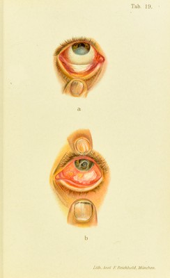
![iittuck ; the ])artici[)atioii of the sclera is more pronounced or a true scleritis develops; and the corneal haze is not so diffuse, but appears in patches, while the vascular changes in tlie superficial and deep layers are more irregular.” The ultimate fate of the corneal maculae varies widely. In some cases transparency is completely regained ; in others, marked by fre(iuent relapses, the center is irre- mediably obscured by tine, nebular opacities and vision is ])ermanently injured. In almost every case of inter- stitial keratitis minute vessels can be detected with a loupe and lateral illumination, or with the ophthalmoscope and direct light. They can be seen twenty years after an attack, and may be utilized as a diagnostic sign of sy])hilis. The cause in two-thirds of the cases is hereditary syphilis, and the classical signs of this condition should always be looked for ; they are : Flat upper jaws, sunken nasal bridge, scars at the anglesof the mouth, and Hutchin- son’s teeth, characterized by diminished size with fairly good enamel and shapely outline, and, usually, wide inter- vals, especially between the incisors. The central incisors of the upper jaw are wedge-shaped at the expense of the free cutting-surface, which is often marked with a small circular notcli. The signitieance of the deformity is limited to the permanent teeth. We also look for ulcers or scars in the palate, for tissue-destruction of the pillars of the fauces, or adhesions of these structures to the pharyngeal wall. Deafness not infrecpiently develops in the later stages of the disease ; the cervical glands are en- larged ; there are chronic periostitis of the tibiae and pain- less synovial effusions in the knee-joint. Upon inquiring into the fiimily history Ave learn of a large mortality among the children, and of abortions and stillt3orn infants. In some cases, if the refracting media are not too much obscured by the opacity, it may be possible, during the liealing-stage, to make out minute light or dark blotches on the eyeground, which I have illustrated in my of OpJithahnoscojyif, vol. vii. of this series, and which I con- sider positive signs of hereditary lues. The larger foci of](https://iiif.wellcomecollection.org/image/b21691587_0184.jp2/full/800%2C/0/default.jpg)