Atlas of the external diseases of the eye : including a brief treatise on the pathology and treatment / by O. Haab ; Authorized translation from the German, edited by G.E. de Schweinitz.
- Haab O. (Otto), 1850-1931.
- Date:
- 1899
Licence: Public Domain Mark
Credit: Atlas of the external diseases of the eye : including a brief treatise on the pathology and treatment / by O. Haab ; Authorized translation from the German, edited by G.E. de Schweinitz. Source: Wellcome Collection.
Provider: This material has been provided by the Royal College of Physicians of Edinburgh. The original may be consulted at the Royal College of Physicians of Edinburgh.
186/312 (page 148)
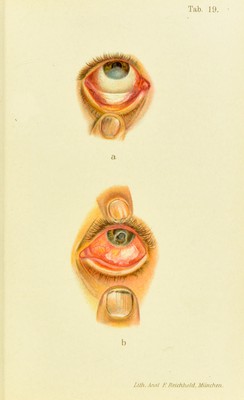
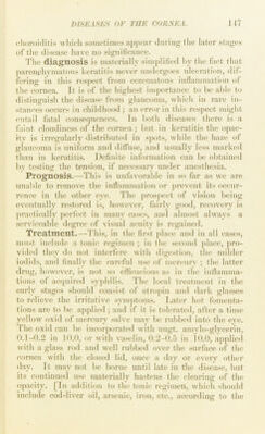
![Plate 20. Herpes zoster ophthalmicus, taken on the sixth daj’^ of the disease. The patient, a healthy man, of 48 years, at that time complained of jjaiii and the sensation of a foreign body in the left eye. The next day he had a slight chill, followed by nausea and lassitude, so that he went to bed. When he woke up the following morning forehead and nose were covered with an eruption which caused burning pain. The left eye also became violently iiidamed and he could not see well with it. The doc- tor ordered lead-water comi)resses (a mistake, on account of the corneal attection), whereupon vision became worse. At the time of admission the vesicles had already dried up and formed crusts, as seen in the pict- ure. The lids are somewhat edematous; conjunctiva very red and swollen, and covered with secretion ; the entire cornea, with the excep- tion of the periphery, is denuded of epithelium, and where any exists it is grayish-white and opaque. The corneal tissue shows diliuse tur- bidity, and the pupil, which is moderately dilated, is barely visible. Sensibility is diminished in the distribution of the ophthalmic branch of the fifth nerve and entirely lost in the cornea except at the periphery. Under a bandage the corneal epithelium gradually regenerated in two weeks and the surface cleared somewliat. When the patient was dis- missed, six weeks after the begiimiiig of the attack, sensibility had not been restored in the cornea; the surface was uneven, though cai)able of reflection, but the tissues were obscured by maculfe, so that the pupil was barely visible. In this case the cornea was attacked primarily, at the same time as the skin. indication.s, suitable diet, regular exerei.se, ma.ssage, etc., the Editor has much faith in the daily inunction of mer- curial ointment, which may be kept up for weeks at a time.] In rare instances ])arenchymatous keratitis is met with in acquired syphilis, usually in a.ssociation with iritis. It is somewhat more common in rheumatic subjects, forming in such ca.ses part of a general scleritis. Portions of the cornea near the .scleritic focus become opaque and, in the course of time, as white as the .sclera {rclerotizing hera- titi.s); the cornea loses its circular outline and appears to be encroached upon by the sclera (Plate 29, h). [The disease is also attributed to rachitis, scrofula, malaria, and depressed nutrition. Parely it may begin in utero.—Ed.] Slight injuries may give rise to extensive parenchyma-](https://iiif.wellcomecollection.org/image/b21691587_0186.jp2/full/800%2C/0/default.jpg)