Atlas of the external diseases of the eye : including a brief treatise on the pathology and treatment / by O. Haab ; Authorized translation from the German, edited by G.E. de Schweinitz.
- Haab O. (Otto), 1850-1931.
- Date:
- 1899
Licence: Public Domain Mark
Credit: Atlas of the external diseases of the eye : including a brief treatise on the pathology and treatment / by O. Haab ; Authorized translation from the German, edited by G.E. de Schweinitz. Source: Wellcome Collection.
Provider: This material has been provided by the Royal College of Physicians of Edinburgh. The original may be consulted at the Royal College of Physicians of Edinburgh.
189/312 (page 149)
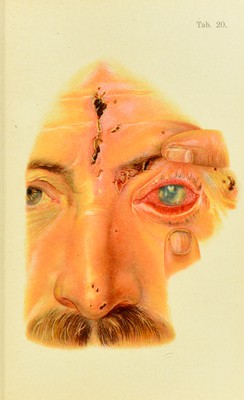
![tons infiltration of the cornea, Avhicli nsnally disappears rapidly, but occasionally jicrsists for some time and only ])artiaily disai)pcars. It is therefore important to observe the greatest care in the treatment of slight injuries, either from scratches or the entrance of foreign bodies; often it may be necessary to use protecting bandages. Deep dif- fuse infiltration of the cornea may also be caused by iridocyclitis. B. CIRCUMSCRIBED INFLAMMATIONS OF THE CORNEA. These forms are more frequent than the diffnse, and the most frequent of them is 2. Eczematous Keratitis, also called phh/ctenular or scrofulous ko-atitis []dilycten- nlar keratoconjunctivitis.] The corneal affection may occur independently or in combination with eczema of the conjunctiva, the predisposing causes being the same for both forms. The pustules vary quite as much in size and number in the independent corneal disease as in conjunctival eczema; but here also each individual focus has a dis- tinctly circular contour. The smaller vesicles, which appear as minute grayish elevations and are rapidly con- verted into small, superficial depressions by the loss of their epithelial covering, heal in from eight to ten days, without causing congestion or leaving any appreciable permanent opacity. The healing of larger idcers takes place much more slowly and involves a greater loss of substance; ulcers with purulent floors are formed; a few thickened blood-vessels appear at the edge of the cornea and gradually work their way toward the ulcer, under- neath the epithelium. Unless secondary infection takes place, the ulcer clears up and regenerates under its fresh ejhthelial covering, as may be seen by its reflective proj)- erties and failure to stain with fluorescin. The normal transparency is not completely restored, but a permanent](https://iiif.wellcomecollection.org/image/b21691587_0189.jp2/full/800%2C/0/default.jpg)