Atlas of the external diseases of the eye : including a brief treatise on the pathology and treatment / by O. Haab ; Authorized translation from the German, edited by G.E. de Schweinitz.
- Haab O. (Otto), 1850-1931.
- Date:
- 1899
Licence: Public Domain Mark
Credit: Atlas of the external diseases of the eye : including a brief treatise on the pathology and treatment / by O. Haab ; Authorized translation from the German, edited by G.E. de Schweinitz. Source: Wellcome Collection.
Provider: This material has been provided by the Royal College of Physicians of Edinburgh. The original may be consulted at the Royal College of Physicians of Edinburgh.
190/312 (page 150)
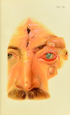
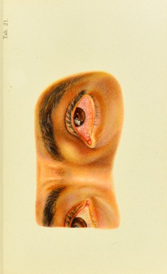
![Plate 21. Foreign Body on the Cornea and Dermoid Cyst of the Orbit.—An Italian inarl)le-cnttcr, 18 years old, yesterday received a splinter in his left eye which appears as a small brown particle surrounded by a yellow infiltra- tion, a little to the temi)oral side of the center. Patient refu.ses to have the dermoid cyst removed. The swelling above the left lachrymal sac has existed since childhood, and has increased very little in the last few years. macul(( rcMiiain.s, es|)eci:illy :ifter a centrally situated ulcer (Plate 2d, h); the circailar shape indicates its cczeiufitous origin. Large pustules may penetrate deeply into the corneal ti.ssne and eventmdly cause a perforation, nsnallv after the development of iritis and turbidity in the anterior chambei’. L:irge single ulcers near the corneal margin are more a[)t to perforate than central ulcers. Perforation is usually followed by attachment of the iris to the wound, where it becomes iiuiarcerated in the heal- ing process (Plate 23, o). If the perforation is very large the iris is apt to .slip through the opening [prolapse of the iris), and if there is extensive purulent infiltration from .secondary infection,' the corneal tissues may break down and a staphyloma result. This is formed as follows; The iris which closes the perforation, although reinforced by granulation- and scar-tissue, is unable to withstand the intraocular pressure, which is usually increased by .secondary glaucoma, and gradiudly bulges forward. In a few'weeks or months the staphyloma is completed—a hemispherical, grayish-white or bluish ])rotrusion, which caiuses a marked deformity. Vision is usually destroyed much earlier. If the disease is protracted and the eruptions constantly recur, accompanied by vascularization, so-called eczematous or scrofulous jjannms (Plate 22) results. Numerous sujjer- ficial blood-vessels unite with new and old foci and their maculfe to form a grayish-red coating over the foce of the cornea and, of conr.se, interfere greatly with vision. If the condition persists for any length of time an extensive](https://iiif.wellcomecollection.org/image/b21691587_0190.jp2/full/800%2C/0/default.jpg)