Atlas of the external diseases of the eye : including a brief treatise on the pathology and treatment / by O. Haab ; Authorized translation from the German, edited by G.E. de Schweinitz.
- Haab O. (Otto), 1850-1931.
- Date:
- 1899
Licence: Public Domain Mark
Credit: Atlas of the external diseases of the eye : including a brief treatise on the pathology and treatment / by O. Haab ; Authorized translation from the German, edited by G.E. de Schweinitz. Source: Wellcome Collection.
Provider: This material has been provided by the Royal College of Physicians of Edinburgh. The original may be consulted at the Royal College of Physicians of Edinburgh.
193/312 (page 151)
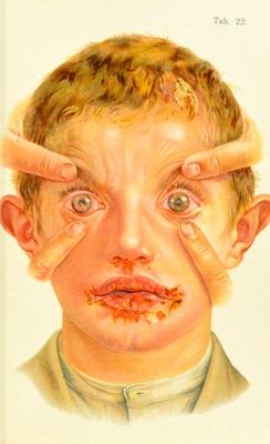
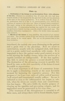
![opacity inav result and cause permanent diminution of visual acuity. Corneal eczema very (d’teii occurs secondarily to eczema of the conjunctiva. Pustules appear directly on the cor- neoscleral junction, ]')artly on the cornea and partly on the conjunctiva. The adjacent corneal area becomes cloudy, and a few blood-vessels make their appearance. This is the so-called marginal Irratifis. Tf the marginal phlyctenulfB are large (1.5—2 mm.) the corneal half is often converted into a deep (excavated) ulcer, with strong ten- dency to perforation (Plate 23, o) ; or the phlyctenular ulcer may leave the ])eripherv and creep toward the cen- ter of the cornea, forming the so-called migratory pustule OY fascicular keratitis. Tlie mechanism of this process is not well understood. Most cases do not come under ob- servation until after the process is completed, weeks or months after the beginning of the inflammation, when the following picture is seen : A bundle of minute blood- vessels, from 1 to 2 mm. broad, extends from some portion of the periphery toward the center of the cornea, running beneath the surface in a straight or slightly curved line and t(‘rminating in a crescentic, grayish elevation. When the j)roce.<?s is kept under observation for some time the blood-vessels a]>pear to ])ush before them the crescentic in- filtration in which they end; the latter gradually wanders toward the center or across the face of the cornea, between the center and the ])eriphery, its convex border presenting toward the center. The process is attended with severe irritation and blepharospasm; children evince a con- stant desire to bury their heads iu pillows or creep into dark corners. Whenever the disease is seen at its inception, the original cause is always found to be a mar- ginal jnistule. Occasionally several fasciculse are seen in the same cornea, or one in each eye. The entire course of the fascicular keratitis across the cornea is marked by a stripe-like opacity, v'hich remains for years as evidence of the disease, and usually ]n’oduces permanent diminution of vision, as it ])referably affects the pupillary region.](https://iiif.wellcomecollection.org/image/b21691587_0193.jp2/full/800%2C/0/default.jpg)