Atlas of the external diseases of the eye : including a brief treatise on the pathology and treatment / by O. Haab ; Authorized translation from the German, edited by G.E. de Schweinitz.
- Haab O. (Otto), 1850-1931.
- Date:
- 1899
Licence: Public Domain Mark
Credit: Atlas of the external diseases of the eye : including a brief treatise on the pathology and treatment / by O. Haab ; Authorized translation from the German, edited by G.E. de Schweinitz. Source: Wellcome Collection.
Provider: This material has been provided by the Royal College of Physicians of Edinburgh. The original may be consulted at the Royal College of Physicians of Edinburgh.
202/312 (page 156)
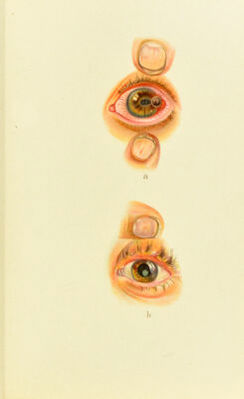
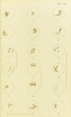
![Plate 24. Herpes Cornese Febrilis.—These eighteen outline drawings show various sliapes and positions of herpes-ulcers, taken from thirteen ciises under my observation during the epidemic of influenza in 1890-91. (They are also to be found in the work of Dr. Hagnauer, wlio was at that time my assistant.') In Figs. 1, 5, 6, 7, 13, and 17 we see macules from former attacks of herpes, recognized by their irregular outline, re- sembling a geographical map. Figs. 10 and 1.5 show the macules which resulted from the ulcers seen in 7 and in 9 and 12. Figs. 12 and 14 illustrate the coalescence of a discrete eruption, and Fig. 17 a magnified jiicture of the original ulcer. In Figs. 11 and 12 slight vascularization is beginning to show itself. even when anesthesia is the only syni])tom, and is impera- tively demanded when there is destruction of the corneal ti.ssne. Herpes cornese febrilis (Horner) .should engage our intere.st if only because it proves conclusively that proc- esses can occur in the cornea in every respect analogous to cutaneous di.sease.s, and that the individual foci po.ssess the .same shape, 011 a very much reduced scale, as in the skin. It is important to note that the vesicles on the cornea are much more delicate in structure, and therefore break down and disappear more rapidly than on the skin; hence the diagnosis is usually made from the characteri.stic shape of the subsequent loss of substance, the herpetic ulcer and its peculiarities. After the vesicles burst (in from one to two days) the cornea for the next week or two looks as if it had been scratched with a sharp sjilinter. The irritation is moderate, and the injured spot as well as its immediate siirrouudings, only slightly opaque. In a week, or from one to two weeks after the hrst a])])earance of the eruption, the last shreds of the vesicle-walls separate and the ulcers present their typical sinuom outline and clear- cut edges. The contour cau be very clearly brought out by framing v'ifli fluorescin, a procedure of the greatest ' Die Misdeiitungen des Herpes Cornese Febrilis, Inaug. Dissert. Zurich, 1891.](https://iiif.wellcomecollection.org/image/b21691587_0202.jp2/full/800%2C/0/default.jpg)