Atlas of the external diseases of the eye : including a brief treatise on the pathology and treatment / by O. Haab ; Authorized translation from the German, edited by G.E. de Schweinitz.
- Haab O. (Otto), 1850-1931.
- Date:
- 1899
Licence: Public Domain Mark
Credit: Atlas of the external diseases of the eye : including a brief treatise on the pathology and treatment / by O. Haab ; Authorized translation from the German, edited by G.E. de Schweinitz. Source: Wellcome Collection.
Provider: This material has been provided by the Royal College of Physicians of Edinburgh. The original may be consulted at the Royal College of Physicians of Edinburgh.
234/312 (page 176)
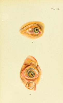
![Plate 30, a. Sarcoma of the Iris.—I am indebted for this picture to the kind- ness of my colleague, Dr. Mayweg, of Hagen, who made the following report of the case before the Ophthalmologic Society of Heidelberg, at the meeting of the society in 1897. As long ago as 1870 the patient, a factory-hand, 53 years old, had his attention called to a small yellowish- brown elevation, as large as a pinhead, in the temporal third of the iris, near the outer rim. A year later he noticed a change in the spot; it gradually became larger, without causing diminution of vision, which at the time of his admission was perfectly normal. The tumor was com- pletely removed in two sittings and the wound healed in three W’eeks. Examination of the tumor revealed a moderately pigmented, spindle- cell sarcoma. The pigment-spots on the lower portion of the iris are not pathologic ; such spots are often seen on the iris; occasionally, however, they develop into sarcoma. h. Syphilitic Iritis.—There is marked ciliary congestion. The hyper- emia in the iris has changed the original blue-gray color (as seen in the other eye in Fig. (J) to a greenish tint. The pupil is dilated with atropin and shows the projecting synechiaj. The patient complains of pain and a moderate degree of photophobia; he was infected a few years ago. removed through an ineision in tlie cornea. AVhen the eye is brought near the electromagnet, if tliere is a loose ])iece of iron in any part of the vitreous body it will be drawn around the lens against the iris and cause it to bulge forward. As soon as this is observed the patient’s head is at once thrown back, or the electric current inter- rupted, lest the substance become fixed in the posterior surface of the iris. If the eye is now turned toward the site of the intruder, it can usually be drawn through the pupillary ojiening into the anterior chamber, if the ])re- caution has been taken to dilate the ])upil before beginning the operation. If the substance is lodged in the retina or choroid coat, some time may elapse before the magnet becomes effective, hence the attempt mu.st not be aban- doned too soon and may be re^ieated if necessary. The smaller the particle of iron the greater must be the power of the magnet, and vice versa. If there is no large mag- net at hand, an attempt must be made to find and extract the foreign body with the small magnetic probe, either through the original scleral wound, or through a fresh in-](https://iiif.wellcomecollection.org/image/b21691587_0234.jp2/full/800%2C/0/default.jpg)