Atlas of the external diseases of the eye : including a brief treatise on the pathology and treatment / by O. Haab ; Authorized translation from the German, edited by G.E. de Schweinitz.
- Haab O. (Otto), 1850-1931.
- Date:
- 1899
Licence: Public Domain Mark
Credit: Atlas of the external diseases of the eye : including a brief treatise on the pathology and treatment / by O. Haab ; Authorized translation from the German, edited by G.E. de Schweinitz. Source: Wellcome Collection.
Provider: This material has been provided by the Royal College of Physicians of Edinburgh. The original may be consulted at the Royal College of Physicians of Edinburgh.
237/312 (page 177)
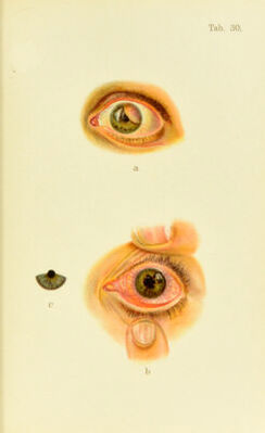
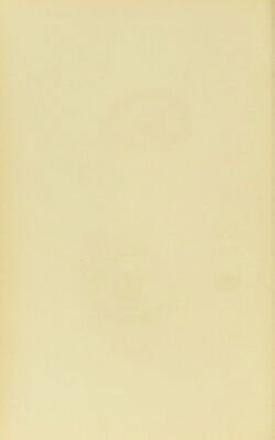
![oision. [If the foreign substance, steel or iron, is [)ro[)- erlv located by means of skiagrapliic examination, the Editor believes that its extraction through a suitably placed ineision by means of the extension point of a Hirschberg or similar magnet is an eminently proper surgical pro- cedure.] DISEASES OF THE IRIS AND CILIARY BODY. I. Inflammation. As a ride, the anterior segments of the uvea, the iris, and the ciliary body uH ])articipate in the inflammatory process, so that inflammation of the iris alone {iritis) or of the ciliary body {ciiciitis) is not very common ; in severe grades of inflammation even the choroid becomes involved, and we speak of uveitis. The symptoms of iritis are very charaeteristic. There is pericorneal injeetion, and the pain, lachrymation, and photophobia are so severe that the patient finds it diflicult or impossible to o})cn his eyes in a bright light. Hyjier- emia is a conspicuous symptom, which manifests itself in chromatic alterations in the iris ; a blue iris becomes green, gray changes to a reddish tint, a light broAvn or green color is somewhat darker and more muddy than that of the normal eye (Plate 30, h). The striationsare partialIv obscured ; the tissues are somewhat turbid and puckered or thickened from the inflammatory infiltration. The in- crease in the volume of the iris causes shrinking and par- tial loss of mobility in the pupil. The latter svm])tom is aggravated by the inflammatory irritation in tiic muscle- fibers, and eventually the ])upil fails to react promptly to light on account of attachments between the pupillary margin and the lens capsule. At first certain portions only of the pupillary margin become attached, which, after dilatation of the pnj)il with a mydriatic (atropin, hyoscin, cocain, homatropin), ajipear as tongue-shaped 12](https://iiif.wellcomecollection.org/image/b21691587_0237.jp2/full/800%2C/0/default.jpg)