Atlas of the external diseases of the eye : including a brief treatise on the pathology and treatment / by O. Haab ; Authorized translation from the German, edited by G.E. de Schweinitz.
- Haab O. (Otto), 1850-1931.
- Date:
- 1899
Licence: Public Domain Mark
Credit: Atlas of the external diseases of the eye : including a brief treatise on the pathology and treatment / by O. Haab ; Authorized translation from the German, edited by G.E. de Schweinitz. Source: Wellcome Collection.
Provider: This material has been provided by the Royal College of Physicians of Edinburgh. The original may be consulted at the Royal College of Physicians of Edinburgh.
241/312 (page 181)
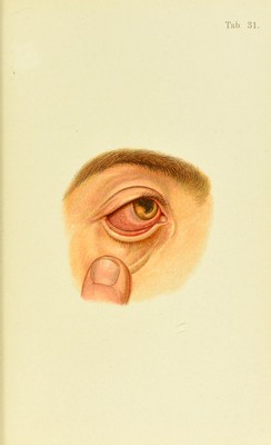
![chronic form must be expressed in months or years, and , the bnal result is but too often practical or total blindness, the more so if the disease shows a tendency to relapses, as it often does. (Mironic iritis and iridocyclitis eventually lead to atrophy of the iris, cataract, atro[)hy of the bulb, either of its anterior portion (j)hfhisis anterior) or of its entire volume [phtlmh buJhi). The causes of iritis and iridocyclitis are manifold. We distinguish between piimary and secondary iritis. The latter occurs in keratitis, especially in the purulent form, and after scleritis, choroiditis, retinal detachments, intraocular tumors, etc. The primary form may follow in the train of various constitutional diseases, or it may occur as a localized dis- order, as in traumatism. Syphilis is by far the common- est cause of iritis and iridocyclitis (in about one-half of all the cases). SypJdlitic iritis may assume the form of a sim])le iritis with synechise (Plate 30, b); or it may be attended with more or less extensive deposits on the cornea, or both these forms may be combined. Not uncommonly the disease presents a characteristic picture by the formation of nod- ules in the tissues of the iris (Plate 31). The color of these nodules when they are small (1 mm.) is grayish-red; when they are large (3 or 4 mm.) yellowish-red, and the surrounding portion of the iris is usually congested and reddened. They preferably select the pupillary, more rarely the ciliary, margin, and grow quite rapidly. Occa- sionally several appear together, and by coalescing convert portions of the pupillary margin into a thick fold. The site of such a syphiloma is invariably marked by a syn- echia, Avhich persists even after the nodules have become absorbed. Any marked thickening of the pu])illary mar- gin, associated with an extensive synechife, should arouse a suspicion of the specific nature of the inflammation, even if the nodules are not plainly visible, as they often require the microscope for their detection. Iritis, with or without the formation of nodules, usually](https://iiif.wellcomecollection.org/image/b21691587_0241.jp2/full/800%2C/0/default.jpg)