Atlas of the external diseases of the eye : including a brief treatise on the pathology and treatment / by O. Haab ; Authorized translation from the German, edited by G.E. de Schweinitz.
- Haab O. (Otto), 1850-1931.
- Date:
- 1899
Licence: Public Domain Mark
Credit: Atlas of the external diseases of the eye : including a brief treatise on the pathology and treatment / by O. Haab ; Authorized translation from the German, edited by G.E. de Schweinitz. Source: Wellcome Collection.
Provider: This material has been provided by the Royal College of Physicians of Edinburgh. The original may be consulted at the Royal College of Physicians of Edinburgh.
259/312 (page 193)
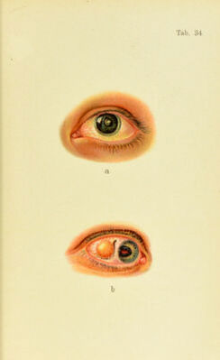
![cases short streaks are seen nearer the peri])liery (Plate 33,1)). Occasionally we meet with a case in which the Ihmellar. cataract remains rnclimentary and the disks are represented by fine disseminated dots. The visual dis- turbance is, of course, correspondingly slight, while in severe cases it is extreme, es])ccially when the pupil is contracted, not a ray of light being able to get past the cataract and reach the retina. Lamellar cataract is usually bilateral and stotionarv, but later in life mav become complete. In the treatment of the first-named varieties of cata- ract operative interference is indicated only when the cata- ract is extensive; lamellar cataract, however, usually requires operative removal sooner or later. Simple iridec- tomy (at the lower, inner angle) may allow some light to enter past the cataract in mild cases ; but, as a rule, removal of the lens by discission is indicated. h. Progressive Cataracts. This group comprises the more frequent forms of cata- ract, the most imjiortant of which is I. Senile Cataract.—It begins as a spoke-like ar- rangement of lines, streaks, or wedges, radiating from the pole of the lens, Inelpieni cataract, goes on developing as caturacta })ituma^ccrir, and becomes complete, c(daracta matnra, when the opacity reaches the capsule, so that the iris does not throw a shadow on the opacity by lateral illumination (Plate 32, a). The cataract is called “rii)c” at this stage, because it is in the best condition for opera- tive removal, the soft consistency of the cortex permitting its complete separation from the ea])sule. In a strong light the yellowish nucleus may be seen shining through the grayish cortical layers. The nucleus may be colorless. Senile cataract is caused by the j)hvsiologic sclerosis of the lens due to age, which is the cause of diminished accom- modation (see p. 53) and serves to protect the nucleus from cataractous change, as shown by the fact that it retains its 13](https://iiif.wellcomecollection.org/image/b21691587_0259.jp2/full/800%2C/0/default.jpg)