Atlas of the external diseases of the eye : including a brief treatise on the pathology and treatment / by O. Haab ; Authorized translation from the German, edited by G.E. de Schweinitz.
- Haab O. (Otto), 1850-1931.
- Date:
- 1899
Licence: Public Domain Mark
Credit: Atlas of the external diseases of the eye : including a brief treatise on the pathology and treatment / by O. Haab ; Authorized translation from the German, edited by G.E. de Schweinitz. Source: Wellcome Collection.
Provider: This material has been provided by the Royal College of Physicians of Edinburgh. The original may be consulted at the Royal College of Physicians of Edinburgh.
260/312 (page 194)
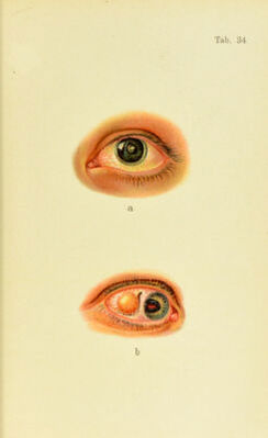
![Plate 34. a. Anterior polar cataract, so-called pyrauiidal cataract. The boj’, who is 15 years old, did not have any purulent discharge at birth; hut six months later he was seized with convulsions. The i)hysician who was called at the time discovered a spot in the eye. Hereditary syi)hilis is suspected, as there were two premature births in the family and eight children died at ages ranging from eight to ten weeks. There are only three children living. No other signs of .syphilis are to be found ; the cornea is perfectly transparent and the fundus of both eyes normal. Visual acuity on both sides is only as the centrally situated opacity, of course, produces marked disturbance, especially with contracted pupil. On both sides a small pointed cone is seen on a round, grayish- white base 2.5 mm. in diameter, projecting into the anterior chamber. The remaining portions of the lens are clear. In performing discission a cir- cular section was made around the polar opacity and the latter extracted. Microscopic examination shows it to be a so-called capsular cataract, situated under the capsule. The operative removal of the lens was successful, and resulted in a visual acuity on both sides of i, with hyper- metropia of 13 D. h. Subconjunctival Displacement of the Lens.—Three months and a half ago the patient, a man of 56 years, struck his eye against the limb of a tree. The accident was immediately followed by marked reduction of visual acuity ; at present the patient is just able to count fingers at 2-i meters. Convex glasses have no efl'ect; the left eye is normal. The scar in the sclera, through which the lens escaped, is plainly seen on the temporal side of the cornea. The pupil is displaced outward, having evidently been turned backward by the force of the blow. Streaks of injected vitreous are seen running toward the seat of rupture. The posterior layers are so turbid with blood that the fundus is barely vis- ible; but a rent in the choroid coat can be seen on the temporal side. The conjunctiva wiis divided and the lens, which had become adherent and annoyed the ])atient, removed without injury to the original rupture or escape of the vitreous. Complete cure in ten days. trans])arency more or less ])erfeetly Avitliin the turbid cortex. After tlie seventietli year tlie sclerosis extends almo.st to the capsule, the usual gray color of the opacity is wanting, and it may ha])])cu that the lens remains .semi- transparent, with a dark reflex due to the absence of corti- cal snb.stance and the yellowish-brown discoloration of the nncleibs—cafaracfa nic/ra. The stage of maturity is followed by that of “ over- ripeness,” during which the volumo ot the lens gradually](https://iiif.wellcomecollection.org/image/b21691587_0260.jp2/full/800%2C/0/default.jpg)