Atlas of the external diseases of the eye : including a brief treatise on the pathology and treatment / by O. Haab ; Authorized translation from the German, edited by G.E. de Schweinitz.
- Haab O. (Otto), 1850-1931.
- Date:
- 1899
Licence: Public Domain Mark
Credit: Atlas of the external diseases of the eye : including a brief treatise on the pathology and treatment / by O. Haab ; Authorized translation from the German, edited by G.E. de Schweinitz. Source: Wellcome Collection.
Provider: This material has been provided by the Royal College of Physicians of Edinburgh. The original may be consulted at the Royal College of Physicians of Edinburgh.
276/312 (page 206)
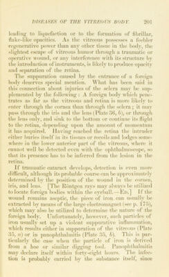
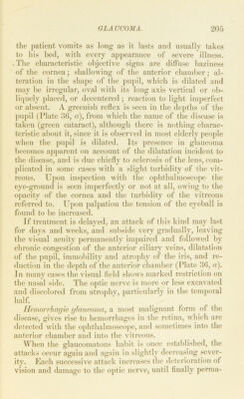
![Plate 36. a. Acute Glaucoma.—E. E., a woman of 71 years, underwent an opera- tion for cataract in the left eye seven years ago (without iridectomy), and since that time enjoyed perfectly good vision, the posterior capsule having been removed by discission shortly after the operation. The pupil was round and movable. Two days ago she was snddenly seized with pain in the eye and dimness of vision, without apparent cause. Her condition improved at first after the administration of myotics, and visual acuity returned to .1. Another acute attack occurred in the clinic, with tension of T i 2, redness of the eye, and dilatation of the pupil (see illustration), which was displaced slightly upward, as frequently happens in glaucoma. The surface of the eyeball was cloudy, and a grayish-green reflex was observed in the deep portions of the eye. Sclerotomy and thorough discission were performed, and by continued use of physostig- miu and pilocarpin the patient was finally cured, with a visual acuity of i. h. Spicule of Iron in the Vitreous (Extracted); Laceration of the Iris, Traumatic Cataract, and Turbidity of the Vitreous.—V. Sch., a peasant- woman, (50 years old, got a piece of iron in her left eye while hoeing potatoes on June 14, 1897. The next day she went to a doctor, who i)io- nouuced the wound superficial and of no consequence. The patient did not feel pain at any time, but comiilaincd of a thick haziness immediately after the injury. When she was admitted to my private hospital, on the 17th, the eye was inflamed and the channel of the wound jdainly seen (see figure). Tlie wound consisted of a rent in the cornea (where the foreign body had entered), a little below tbe center, appearing as a fine, gray line 1..5 mm. in length ; a laceration in the iris, traumatic cataract, and a triangular wound in tbe iiostcrior capsule. The iris was adherent to the wound in the anterior capsule. The pupillary reflex was greenish, and by lateral illumination a metallic Imster was seen in the opacity of the posterior cortex. The foreign body was not visible, although undoubtedly present in the vitreous. On approaching the large electromagnet it was at once drawn into the anterior chamber, from which it was removed through the original point of entrance (June 17). On the 27th the in- flammation had j)ractically disappeared, and on the 29th the woman was discharged. All inflammatory symptoms had subsided and the patient w'as able to count fingers at 2 meters. On July 9 the visual acuity was still tbe same and tbe cataract had not progressed; the fundus also was plainly visible. nent, tilxsoliite blindne.ss sii})ervenes and the eye present.s the ti])pearances (iharacteri.^^tic of absolute glaucomaj The cornea i.s less o])a(pie tlian in the early stages, and is snr- rounded by a wreath of dilated blood-vessels j the anterior](https://iiif.wellcomecollection.org/image/b21691587_0276.jp2/full/800%2C/0/default.jpg)