Atlas of the external diseases of the eye : including a brief treatise on the pathology and treatment / by O. Haab ; Authorized translation from the German, edited by G.E. de Schweinitz.
- Haab O. (Otto), 1850-1931.
- Date:
- 1899
Licence: Public Domain Mark
Credit: Atlas of the external diseases of the eye : including a brief treatise on the pathology and treatment / by O. Haab ; Authorized translation from the German, edited by G.E. de Schweinitz. Source: Wellcome Collection.
Provider: This material has been provided by the Royal College of Physicians of Edinburgh. The original may be consulted at the Royal College of Physicians of Edinburgh.
294/312 (page 216)
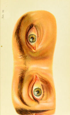
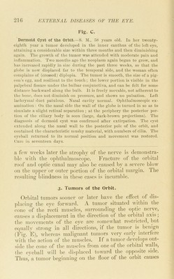
![Fig. C. Dermoid Cyst of the Orhit.—S. M., 58 years old. lu her twenty- eighth year a tumor developed in the inner canthus of the left eye, attaining a considerable size within three months and then diminishing again. The growth of the tumor was attended with moderate pain and inflammation. Two months ago the neoplasm again began to grow, and has increased rapidly in size during the past three weeks, so that the glol)e is now displaced far to the temporal side, and the woman often complains of (crossed) diplopia. Tlie tumor is smooth, the size of a pig- (mn's egg, and resilient to the touch ; the lower portion is visible in the l)alpebral fissure under the bulbar conjunctiva, and can be felt for some distance backward along the bulb. It is freely movable, not adherent to the bone, does not diminish on pressure, and shows no pulsation. Left lachrymal duct patulous. Nasal cavity normal. Ophthalmoscopic ex- amination : On the nasal side the wall of the globe is turned in so as to simulate a slight retinal separation ; at the periphery the posterior por- tion of the ciliary body is seen (large, dark-brown projections). The diagnosis of dermoid cyst was confirmed after extirpation. The cyst extended along the nasal wall to the posterior pole of the orbit, and contained the characteristic mushy material, with numbers of cilia. The eyeball returned to its normal position and movement was restored. Cure in seventeen days. a fow weeks later the atrophy of the nerve is demonstra- ble with the ophthalmoscope. Fracture of the orbital roof and optic canal may also be caused by a severe blow on the upper or outer portion of the orbital margin. The resulting blindness in these cases is incurable. 3. Tumors of the Orbit. Orbital tumors sooner or later have the effect of dis- ])lacing the eye forward. A tumor situated within the cone of the recti mu.scles, surrounding the o])tic nerve, causes a displacement in the direction of the orbital axis; tlie movements of the eye are somewhat restricted, but ecpially strong in all directions, if the tumor is benign (Fig. F), whereas malignant tumors very early interfere with the action of the mu.scles. If a tumor develops out- side the cone of the muscles from one of the orbital vails, the eyeball will be di.sj)laced toward the opposite side. Thus, a tumor beginning on the floor of the orbit causes](https://iiif.wellcomecollection.org/image/b21691587_0294.jp2/full/800%2C/0/default.jpg)