Atlas of the external diseases of the eye : including a brief treatise on the pathology and treatment / by O. Haab ; Authorized translation from the German, edited by G.E. de Schweinitz.
- Haab O. (Otto), 1850-1931.
- Date:
- 1899
Licence: Public Domain Mark
Credit: Atlas of the external diseases of the eye : including a brief treatise on the pathology and treatment / by O. Haab ; Authorized translation from the German, edited by G.E. de Schweinitz. Source: Wellcome Collection.
Provider: This material has been provided by the Royal College of Physicians of Edinburgh. The original may be consulted at the Royal College of Physicians of Edinburgh.
300/312 (page 222)
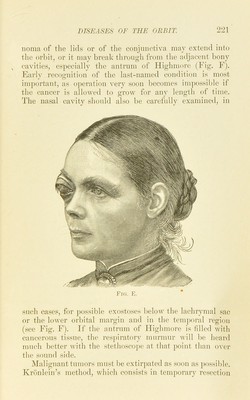
![‘2-29. Fig. F. Cancer of tbe Upper Maxilla and OrPit.—Mrs. A. W., 53 years old. Four mouths before lier first visit to tlie clinic the patient had violent toothache on the right side, and although she had several teeth extracted the pains increased in seveidty and spread to the right ej’e and temi)le. As we see in the picture, there was even at that time a slight swelling in the region of the right upper jaw and temple, and the right eyeball w;is displaced forward and upward. The movements of the eye were restricted in every direction, but especially downward. The patient was referred to the surgical clinic, and e.xtirpation of the tumor was performed by my colleague. Dr. Kroulein. The whole upper jaw, with the exception of part of the palatine process, and the greater part of the zygoma were re- sected. It was then found that the growth was much more extensive than was to be expected ; it extended some distance backward on the base of the skull and was laid bare as far as the middle meningeal artery. The eyeball bad to be removed, as it was in close relation with the can- cerous tissue. Externally the tumor had broken through the bone and invaded the masseter muscle. On microscopic examination it proved to be a squamous epitbelioma. The woman became extremely emaciated and died three months later in collapse. There was no I’ecurrence of the tumor. of the temporal wall of the orbit, affords the best means of access. The flap of bone and soft tissues is replaced after the tumor has been removed and secured with sutures. A small tumor in the temporal portion of the orbit can often be removed by this method without sacrificing the globe. In extensive malignant growths the entire con- tents of the orbit must bo extirpated. An interestini^ and not very common affection of the orbit has received the name pulsating- exophthalmos (Plate 40). ft may develop .spontaneously (rarely) or after a severe blow on the skull. Pulsation can be felt at the inner u])per portion of the bulb, and the patient complains of a noise in his head like the ])ounding of a steam-engine. A bruit can be di.stinctlv heard on au.scul- tation in the region about the eye and more faintly as far back as the occij>ut, on both the affected and the unaffected sides. Ft is a characteri.stic sign that the noise and pidsa- tion di.sappear on com])ression of the carotid. F^pon close inspection a large pulsating vein is usually detected in the](https://iiif.wellcomecollection.org/image/b21691587_0300.jp2/full/800%2C/0/default.jpg)