Volume 1
A text-book of human physiology : including histology and microscopical anatomy : with special reference to the requirements of practical medicine / by L. Landois ; translated from the seventh German edition, with additions, by William Stirling.
- Date:
- 1891
Licence: Public Domain Mark
Credit: A text-book of human physiology : including histology and microscopical anatomy : with special reference to the requirements of practical medicine / by L. Landois ; translated from the seventh German edition, with additions, by William Stirling. Source: Wellcome Collection.
Provider: This material has been provided by the Royal College of Physicians of Edinburgh. The original may be consulted at the Royal College of Physicians of Edinburgh.
107/602 page 67
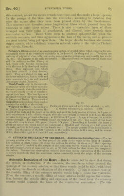
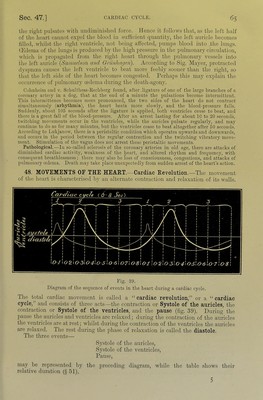
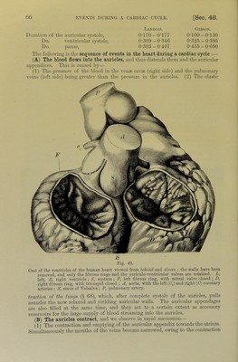
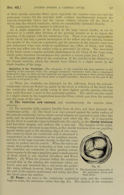
![of thoir circular umscular fibres (more especially the superior vena cava and the pulmonary veins); (2) the auricular u'alls contract simultaneously towards the auriculo-ventricular valves and the venous orifices, whereby (3) the blood is driven into the relaxed ventricles, which arc considerably distended thereby. The contraction of the auricles is followed by— (a) A slight stagnation of the blood in the large venous trunks, as can be observed in a rabbit after division of the pectoral muscles so as to expose the junction of the jugular with the subclavian vein. There is no actual regurgitation of the blood, but only a jiartial interruption of the inflow into the auricles, because the mouths of the veins are contracted, and the pressure in the superior vena cava and pulmonary veins soon holds in equilibrium any reflux of blood; and lastly, because any reflux into the cardiac veins is prevented by valves. The movement of the heart causes a regular pulsatile phenomenon in the blood of the vente cavfe, which under abnormal circumstances may produce a venous pulse (see § 99). (b) The chief motor effect of the contraction of the auricles is the dilaUdion of the relaxed ventricle, Avhich has already been dilated to a slight extent by the elastic traction of the lungs. Aspiration of the Ventricles.—The dilatation of the ventricles has been ascribed to the elasticity of the muscular wails—the strongly contracted ventricular walls (like a compressed india-rubber bag), in virtue of their elasticity, are supposed, in returning to their normal restino' form, to suck in or aspirate the blood under a negative pressure ; this power on the part of the ventricle is not great (p. 68). (c) A^Tlen the ventricles are distended by the inflowing blood, the anriculo- ventricnlar valves are floated up, partly by the recoil or reflexion of the blood from the ventricular wall, and partly owing to their lighter specific gravity, whereby they easily float into a more or less horizontal position. The valves are also raised to a slight extent by the longitudinal muscular fibres, Avhich pass from the auricles into the cusps of the valve. (C) The ventricles now contract, and simultaneously the auricles relax, whereby— (1) The muscular walls contract forcibly from all sides, and thus diminish the ventricular cavity. (2) The blood is at once pressed against the under surface of the auriculo-ventricular valves, Avhose curved margins are opposed to each other like teeth, and are pressed hermetically against each other (fig. 40). It is impossible for the blood to push the cusps backwards into the auricle, as the chordae tendineae hold fast their margins and surfaces like a taut sail. The margins of the neigh- bouring cusps are also kept in apposition, as the chordae tendine^ from one papillary muscle always pass to the adjoining edges of Wo cusps. The extent to which the ventricular Avail is shortened is compensated by the contraction of the pa]hllary muscle, and also of the large muscular chordae, so that the cusps cannot be pushed into the auricle. When the valves are closed, their surfaces are horizontal, so that, even Avhen the vejitricles are contracted to their greatest extent, there remains in the supra-papillarij space a small amount of blood Avhich is not expelled {Sandhorri and Worm Muller). (3) When the pressure Avithin the ventricles e.xceeds that in the arteries, the semi-lunar valves are forced open and stretched like a sail across the jiocket- Eig, 41. hke sinus without, hoAvever, being directly applied to The closed semi-lunar valve ot tlie Avail of the arteries (pulmonary and aorta), and thus the pulmonary artery seen the blood enters the arteries. from below. (L) Pause. As soon as the ventricular contraction ends, and the ventricles begin to relax, the semi-lunar valves close (fig. 41). The diastole of the ventricles](https://iiif.wellcomecollection.org/image/b21981516_0001_0107.jp2/full/800%2C/0/default.jpg)
 No text description is available for this image
 No text description is available for this image
 No text description is available for this image