Volume 1
A text-book of human physiology : including histology and microscopical anatomy : with special reference to the requirements of practical medicine / by L. Landois ; translated from the seventh German edition, with additions, by William Stirling.
- Date:
- 1891
Licence: Public Domain Mark
Credit: A text-book of human physiology : including histology and microscopical anatomy : with special reference to the requirements of practical medicine / by L. Landois ; translated from the seventh German edition, with additions, by William Stirling. Source: Wellcome Collection.
Provider: This material has been provided by the Royal College of Physicians of Edinburgh. The original may be consulted at the Royal College of Physicians of Edinburgh.
133/602 page 93
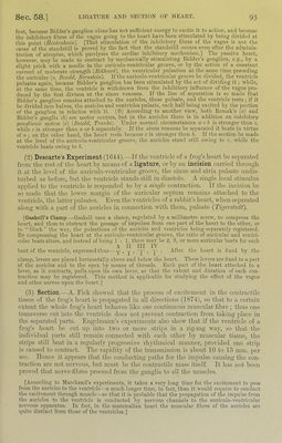
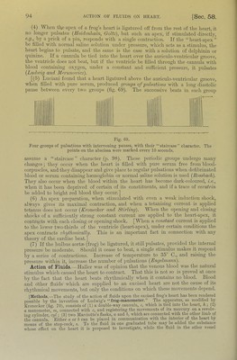
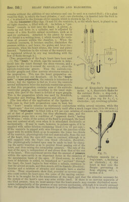
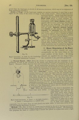
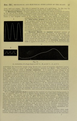
![first, because Bidder’s ganglion alone has not sufficient energy to c.xcite it to action, and because the inhibitory filires of the vagus going to the heart have been stimulated by being divided at this point {Ileklcnhain). [That stimulation of the inliibitory fibres of the vagus is not the cause of the standstill is proved by the fact that the standstill occurs even after the adminis- tration of atropine, which paralyses the cardiac inhibitory mechanism.! 'J'he passive heart, however, maybe made to contract by mechanically stimulating Bidder’s ganglion, e.cj., by a slight prick with a needle in the auriculo-ventricular groove, or by the action of a constant current of moderate strength {Ec.khard), the ventricular ]mlsation at the same time preceding the auricular (v. Bezold, Bermtein). If the auriculo-ventricular groove be divided, the ventricle pulsates again, because Bidder’s ganglion has been stimulated by the act of dividing it; while, at the same time, the ventricle is withdrawn from the inhibitory influence of the vagus pro- duced by the first divison at the sinus venosus. If the line of separation is so made that Bidder’s ganglion remains attached to the auricles, these pulsate, and the ventricle rests ; if it be divided iiRo halves, the auricles and ventricles pulsate, each half being e.xcited by the portion of the ganglion in relation with it. (B) According to another view, both Remak’s (a) and Bidder’s ganglia (h) are motor centres, but in the auricles there is in addition an inhibitory ganglionic sifstem. (c) {Bezold, Tmiibc). Under normal circumstances a + b is stronger than c, while c is stronger than a or b separately. If the sinus venosus be separated it beats in virtue of a ; on the other hand, the heart rests because c is stronger than b. If the section be made at the level of the auriculo-venti'icnlar groove, the auricles stand still owing to c, while the ventricle beats owing to b. (2) Descarte’s Experiment (1644).—If the ventricle of a frog’s heart he separated from tlie rest of the heart by means of a ligature, or liy an incision carried through it at the level of the anricnlo-ventricnlar groove, the sinus and atria pulsate undis- turbed as before, but the ventricle stands still in diastole. A single local stimulus applied to the ventricle is responded to by a smgle contraction. If the incision be so made that the lower margin of the auricular septum remains attached to the ventricle, the latter pulsates. Even the ventricles of a rabbit’s heart, when separated along with a part of the auricles in connection with them, pulsate (Tigerstedt). [GaskeU’s Clamp.—Gaskell uses a clamp, regulated by a millimetre screw, to compress the heart, and thus to obstruct the passage of impulses from one part of the heart to the other, or to “block” the way, the pulsations of the auricles and ventricles being separately registered. By compressing the heart at the auriculo-ventricular groove, the ratio of auricular and ventri- cular beats alters, and instead of being 1 : 1, there may be 2, 3, or more auricular beats for each A II III IV beat of the ventricle, expressed thus—y , j- , -j^ . ^ . After the heart is fixed by the clamp, levers are placed horizontally above and below the heart. These levers are fixed to a part of the auricles and to the apex by means of threads. Each part of the heart attached to a lever, as it contracts, pulls upon its own lever, so that the extent and duration of each con- traction may be registered. This method is applicable for studying the effect of the vagus and other nerves upon the heart.] (3) Section.—A. Eick showed that the process of excitement in the contractile tissue of the frog’s heart is propagated in aU directions (1874), so that to a certain extent the whole frog’s heart behaves like one continuous muscular fibre; thus one transverse cut into the ventricle does not prevent contraction from taking place in the separated parts. Engehnann’s experiments also show that if the ventricle of a frog’s heart be cut u]> into two or more strijis in a zig-zag way, so that the individual parts still remain connected with each other by muscular tissue, the strips still beat in a regularly ju’ogressive rhythmical manner, provided one strip is caused to contract. The rapidity of the transmission is about 10 to 15 mm. per sec. Hence it appears that the conducting paths for the impulse causing the con- traction are not nervous, but must be the contractile mass itself. It has not been proved that nerve-fdires proceed from the ganglia to all the muscles. [According to Marchand’s experiments, it takes a very long time for tlie excitement to jiass from the auricles to the ventricle—a much longer time, in fact, than it would require to conduct the e,xcitpment through muscle—so that it is ]irobable tliat the propagation of the impulse from the auricles to the ventricle is conducted lij' nervous channels to the auriculo-ventricular nervous apparatus. In fact, in the mammalian heart the muscular fibres of the auricles are quite distinct from those of the ventricles.]](https://iiif.wellcomecollection.org/image/b21981516_0001_0133.jp2/full/800%2C/0/default.jpg)
 No text description is available for this image
 No text description is available for this image
 No text description is available for this image