Volume 1
A text-book of human physiology : including histology and microscopical anatomy : with special reference to the requirements of practical medicine / by L. Landois ; translated from the seventh German edition, with additions, by William Stirling.
- Date:
- 1891
Licence: Public Domain Mark
Credit: A text-book of human physiology : including histology and microscopical anatomy : with special reference to the requirements of practical medicine / by L. Landois ; translated from the seventh German edition, with additions, by William Stirling. Source: Wellcome Collection.
Provider: This material has been provided by the Royal College of Physicians of Edinburgh. The original may be consulted at the Royal College of Physicians of Edinburgh.
171/602 page 131
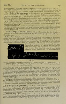
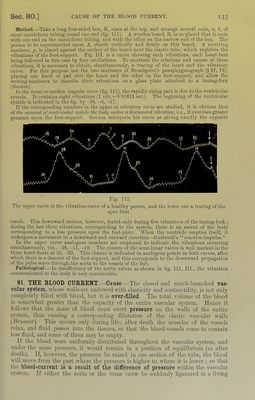
![I3I as 62 continietres) ; carotid and Iomoral = 0’068 second ; femoral (inguinal region) and posterior tibial = 0-097 second (distance estimated at 91 centimetres). [Waller obtained between the heart and carotid 0 10 second ; heart and lemoral, 0T8 sec.; heart and dorsalis pedis, 022.] The velocity of the pulse-wave in the arteries of the upper extremities = 8-43 metros per second, and in those of the lower extremity 9'40 metres per second, \i.e., about 30 feet per second]. The velocity is greater in the less extensile arteries of the lower extremities than in those of the upper limb. For the same reason it is less in the peripheral arteries and in the yielding arteries of children {Czermak). E. H. Weber estimated the velocity at 9-24 metres per second ; Garrod, 9-10'8 metres ; Grashey, 8'5 metres ; Moens, 8‘3 metres, and with diminished pressure during Valsalva's experi- ment 7‘3 metres (§ 60, § 74). ^ Influencing Conditions.—In animals, hamiorrhage, slowing of the heart produced by] stimula- tion of the vagus {Moens), section of the spinal cord, deep morphia-narcosis, and dilatation of the blood-vessels by heat, produce slowing of the velocity, while stimulation of the spinal cord accelerates it (Grunmach). The wave-length of the pulse-wave is obtained by multiplying the duration of the inflow of blood into the aorta = 0’08 to 0'09 second (5 51), by the velocity of the pulse-wave. Method. Place the knobs of two tambours (fig. 88) upon the two arteries to be investigated, 01 place one over the apex-beat aud the other upon an artery. These receiving tambours are connected with t\yo registering tambours, as in Brondgeest's pausphygmograph (§ 67, fig. 88), so that their \yi'iting-levers are dhectly over each other, and so arranged as to write simultane- ou-sly on one vibrating plate attached to a tuning-fork. [Or they may be made to write upon a revolving cylinder, whose rate of movement is ascertained by causing a tuning-fork of a known rate of vibration to wi’ite under them.] The apparatus is improved by using rigid tubes and filling them with water, in which all im- pulses are rapidly communicated. In arteries which are Fig. 109. A, curve of radial artery on a vibrating surface (1 vib. =0-01613 see.) ; P, apex of curve ; e, e, elastic vibrations ; R, dicrotic wave. B, curve of same radial taken along with the heart- beat ; V, H, P, conti-action of the ventricle. distant from each other, or in the case of the heart and an artery, the two knobs of the ^ceiving tambours may be connected by means of a Y-tube with o?ie writing-lever. In fig. 109, B 1.S a curve from the radial artery taken in this way. In it « H P indicates contraction°of the ventricle ; H, the apex of the venti-iciilar contraction ; P, the primary apex of the radial curve ; V, the beginning of the venti-icular contraction ; p, of the radial pulse. A is the curve of the radial artery alone. From these curves it is evident that in this instance nine vibrations occur between the beginning of the ventricular conti-action and the beginning of the pulse in the radial artery = 0-1.5 sec. difference between the carotid and the posterior tibial pulse = 0-137 sec. Bathological._—In cases of diminished externihility of the arteries, e.g., in atheroma (§ 77 D) the pulse-wave is propagated more rapidly. Local dilatations of the arteries, as in aneurisms', caMe a reterdation of the wave, and a similar result arises from local constrictions. Relaxation of tlie walls of the vessels in high fever retards the movement {Hanieinijk). 79. OTHER PULSATILE PHENOMENA.—1. In the mouth and nose, when they are filled with air and the glottis closed, pulsatile phenomena (due to the arteries in their soft parts), may be found communicating a movement to the contained air. The curves obtained are relatively small, and closely resemble the curve of the carotid. A similar pulse is obtained in ttie tympanum with intact membrana tympani, and when the soft parts of the tympanum are congested {Schwartze, Troltsch). 2. Entoptical Pulse.—After violent exercise, an illumination, corresponding to each pulse- beat, occurs on a dark optical field. When the optical field is bright, an analogous darkening occurs. The ophthalmoscope occasionally reveals pulsation of the retinal arteries {Jiiger), which becomes marked in insufficiency of the aortic valves.](https://iiif.wellcomecollection.org/image/b21981516_0001_0171.jp2/full/800%2C/0/default.jpg)
 No text description is available for this image
 No text description is available for this image
 No text description is available for this image