Volume 1
A text-book of human physiology : including histology and microscopical anatomy : with special reference to the requirements of practical medicine / by L. Landois ; translated from the seventh German edition, with additions, by William Stirling.
- Date:
- 1891
Licence: Public Domain Mark
Credit: A text-book of human physiology : including histology and microscopical anatomy : with special reference to the requirements of practical medicine / by L. Landois ; translated from the seventh German edition, with additions, by William Stirling. Source: Wellcome Collection.
Provider: This material has been provided by the Royal College of Physicians of Edinburgh. The original may be consulted at the Royal College of Physicians of Edinburgh.
213/602 page 173
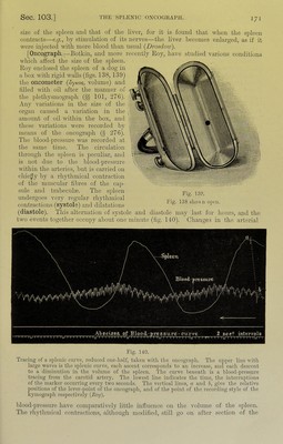
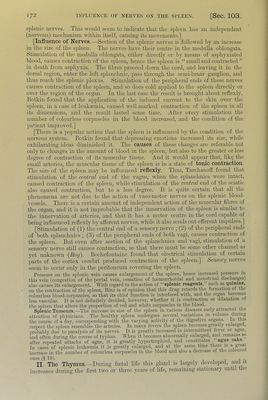
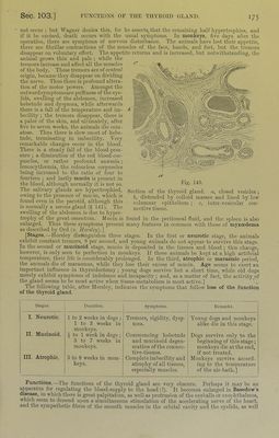
![173 teiitli or fourtoeutli year, when it l)Cgius to atrophy and undergo fatty degeneration. [The degeneration begins at tlie outer part of each lobule and progresses inwards (//<*’). Waldeyer hnds that even in the oldest j)erson the thymus is always repre- sented by a mass of fat, at least as largo as tlie thymus at birth, and always con- taining some adenoid tissue either in a diffuse or nodular form.] Structure.—“It consists of an aggregation of lymph-follicles (resembling the glands of Peyer) or masses of adenoid tissue held together by a frameworlc of connective-tissue which contains blood-vessels, lymphatics, and a few nerves (fig. 141). The framework of connective-tissue gives off septa which divide the gland into lobes, these being further subdivided by finer septa into lobules, the lobules being separated by fine intra-lobular lamellte of connective-tissue into follicles (0‘5- 1'5 mm.). These follicles make up the gland- substance, and they are usually polygonal when seen in a section. Each follicle consists of a cortical and a medullary part, and the matrix or framework of both consists of a fine adenoid reticulum Avhose meshes are filled with lymph- corpuscles” (fig. 142, rt).] Many of these cor- puscles exhibit various stages of disintegration. In the medulla are found the concentric cor- puscles of Hassal. [“They consist of a central gTanular part, around Avhich are dis- jiosed layers of flattened nucleated endothelial cells arranged concentrically. When seen in a section they resemble the ‘ cell-nests ’ of (“pithelioma (fig. 142, b). They have also been occur in the prostate. They are most Fig. 141. Section of the thymns gland of a cat, with one complete lobule with a cortical part a, and a centre, b. a, lymphoid tissue; c, blood-vessels injected; d, connective-tissue. coni]3ared to similar bodies which numerous Avhen the gland undergoes its frog IS undergoing retrograde metamorphosis.” Sig. Mayer finds that the thymus of the contains structures, Avith transverse markings, identical with the stripes of striped muscular fibres. The struc- tures are identical Avith those called “ sarcoplasts ” by Margo and Paneth, and “ sarcolytes ” by fSig. Mayer. They also occur in large numbers in the tail of the larvre of batrachians, Avhen the tail retrograde metamorphosis.] Simon, His, and others described a convoluted blind canal, the “central canal,” as occurring Avithin the gland, and on it the follicles Avere said to be placed. ()ther observers, Jendrassik and Klein, either dejiy its existence or regard it merely as a lymphatic or an arti- ficial product. Kui7ierous fine lymphatics penetrate into the interior of the organ, and many are distributed over its surface, but their mode of origin is unknoAvn. [Tliey seem to bo channels through Avhich the lympli- corpuscles are convoyed aAA'ay from the gland.] Numerous biood-vessels are also distributed to the septa and follicles (fig. 141, c). Chemical Composition.—Besides gelatin, albumin, soda-albumin, there are sugar and fat, leucin, xanthin, hypoxantliin, formic, acetic, butyric, and succinic acids. Potash and phosphoric acid are more abundant in the ash than soda, calcium, magnesium (? ammonium), chlorine, and suljihuric acid. Function of the Thymris.—As long as it exists, it seems to jierform the functions of a true lymph-gland. This view is supported by tlie fact that in reptiles and amphibians, which do not Fig. 142. Elements of the thymus ( X 300). a, lym2)h-cor2)us- cles ; b, concentric corpuscle of H assail.](https://iiif.wellcomecollection.org/image/b21981516_0001_0213.jp2/full/800%2C/0/default.jpg)
 No text description is available for this image
 No text description is available for this image
 No text description is available for this image