Volume 1
A text-book of human physiology : including histology and microscopical anatomy : with special reference to the requirements of practical medicine / by L. Landois ; translated from the seventh German edition, with additions, by William Stirling.
- Date:
- 1891
Licence: Public Domain Mark
Credit: A text-book of human physiology : including histology and microscopical anatomy : with special reference to the requirements of practical medicine / by L. Landois ; translated from the seventh German edition, with additions, by William Stirling. Source: Wellcome Collection.
Provider: This material has been provided by the Royal College of Physicians of Edinburgh. The original may be consulted at the Royal College of Physicians of Edinburgh.
214/602 page 174
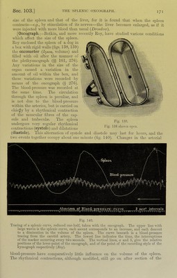
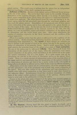
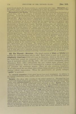
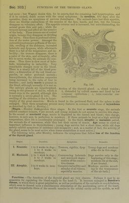
![possess lymph-glands, the thymus remains as a permanently active organ. [Extirpation gave lew positive results, but chemioal investigation shows that the parenchyma contains a large number of products indicating considerable metabolic activity (FriccUcbcn).] [Development of the Thymus.—The thymns is the organ which earliest shows the structure of adenoid tissue, both in the ontogeny of individual mammals and in the jiliylogeny of the vertebrates. In man His maintains that it is derived from the epithelium covering the fourth, third, and part of the second branchial cleft, which becomes compressed in the angle between the head and neck. (More recent observers—Ivastochenko and others—have thrown some doubt on the correctness of His’s observations; jirobably there are considerable dilferences in dilferent classes of vertebrates, but all are now agreed that the original thymns is an epithelial organ mainly derived from the epithelinm covering the gill-clefts.) The tube of epithelium—sinus prfecervicalis—so formed grows inwards, branching dichotomonsly, and ramifying in the connective-tissue behind the sternum, jnst above the pericardium. The cells forming it gi'ow inwards and till np the lumen of the “gland,” and at last of the duct also. By this epithelial ingrowth the same condensation of connective-tissue is brought about as in the tonsil, and in the same way blood-vessels appear in large nnmbei-s, and leucocytes begin to wander out of the vessels, are detained in the meshes of the connective-tissue, aud invade the nearly functionless epithelial gland lobules. The cells of the latter proliferate, and the older cells of the lobnle are pushed to the centre, become cornified, and present very much the appearance of the cell-nests of an epithelioma, forming the so-called “concentric corpuscles of Hassall.” While the leuco- cytes soon eat away the majority of the epithelial cells, and break the continuity of the e]iithelial tubes, these cornified sti’uctures long resist their attacks, and the thymus always retains the lobular character imparted to it by its epithelial precursor. The leucocytes divide rapidly by mitosis in the connective-tissue surrounding these epithelial remains, though no ti-ue “germ-centres” are ever formed. The complete removal of the “concentric corpuscles” by the leucocytes leads to the disappearance of the latter, and the appearance of fat in the position of the thymus ; but Waldeyer has recently shown that the outward form of the thymus is always preserved in this fatty mass, and that it is always possible to demonsteate microscopi- cally in some yiart of it a remaining leucocyte infiltration, and wherever this is at all well marked it will be found to surround a surviving “concentric corpuscle” ((?. L. Cbulland).] III. The Thyroid.—Structure.—The gland consists of lobes and lobules held together by connective-tissue rich in cells. Each lobule is made up of numerous completely closed sacs (0-04 toOT mm. in diameter), which in the embryo and the newly-born animal are composed of a membrana propria lined by a single layer of nucleated cubical cells (fig. 143). The sacs contain a transparent, viscid, albuminous fluid, [blot unfrequently the sacs contain many coloured Wood- corpuscles (jBaie?').] Each sac is surrounded hy a plexus of capillaries which do not penetrate the membrana propria. There _ are also numerous lymphatics. At an early period the sacs dilate, their cellular luring atrophies, and their contents undergo colloid degeneration. When the gland-vesicles are greatly enlarged, “goitre ” is produced. The chemical composition of this gland has not been much investigated. In addition to the ordinary constituents, leucin, xanthin, saidtin, lactic, succinic, and volatile fatty acids have ^^[Ldsion.—The effects differ according to the animal operated on. This gland l)as been exUed in the human subject in cases of g.ntre. Reverdin pointed out that a peculiai conditio results called cachexia stumipriva, and practically the human being becomes a cictiik T s opemton the^ is highly questionable when performed on man. Rabbits endure ti e operation well, and so do the sheep, calf, and horse, none of the remarkable symptoms that oLur in the dog and monkey being manifested by them. In pigeons no obvious disturbance r. produced after bilateral excision of these glands, so that they do not Lt function in these animals {R. Eioald). Of dogs, cats, and foxes, survive ; nearly all die. It appears therefore that herbivora bear the opeiation and ^'‘“^1 [ew e after-effects tliL carnivora {Sa-nqicirico and Orecchia). I he immediate efiects a, e hbiillai conti-actions which ultimately influence the gait of the animals, convulsions, anresthcsia, ^eat Zinutiorof S losi of fle.sh, redness of the ears, and intense heat of the skin (wind disannear after several days), difficulty in seining and eating food, keiato-conjunctn itis, and r llnflv flisturbaiice of the rhythin of respiration with dyspiicea and spasms of the abdominal](https://iiif.wellcomecollection.org/image/b21981516_0001_0214.jp2/full/800%2C/0/default.jpg)
 No text description is available for this image
 No text description is available for this image
 No text description is available for this image