Volume 1
A text-book of human physiology : including histology and microscopical anatomy : with special reference to the requirements of practical medicine / by L. Landois ; translated from the seventh German edition, with additions, by William Stirling.
- Date:
- 1891
Licence: Public Domain Mark
Credit: A text-book of human physiology : including histology and microscopical anatomy : with special reference to the requirements of practical medicine / by L. Landois ; translated from the seventh German edition, with additions, by William Stirling. Source: Wellcome Collection.
Provider: This material has been provided by the Royal College of Physicians of Edinburgh. The original may be consulted at the Royal College of Physicians of Edinburgh.
220/602 page 180
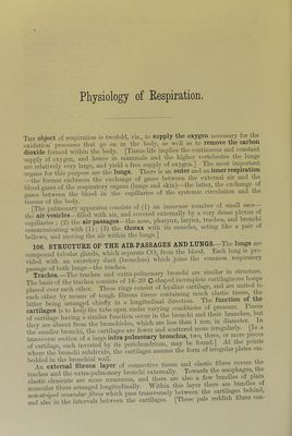
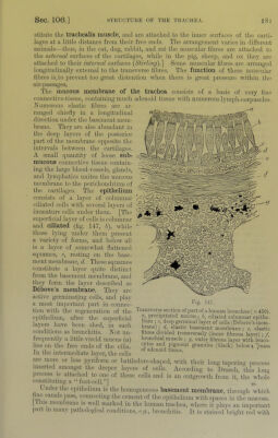
![Physiology of Eespiration. The obiect of respiration is twofold, viz., to supply the oxygen necessary for the oxidation processes that go on in the body, as well as to remove the carbon dioxide formed within the body. [Tissuedife implies the contmuous and constant supply of oxygen, and hence in mammals and the higher vertebrates the lungs are relatively very large, and yield a free supply of oxygen.] The most importmt organs for this purpose are the lungs. There is an outer and an inner respiration —the former embraces the exchange of gases between the external air and the blood-gases of the respiratory organs (lungs and skin)—the latter, the exchanp oi gases between the blood in the capiharies of the systemic circidation and the tissues of the body. . ii [The pulmonary apparatus consists of (1) an immense number of small sacs the air-vesicles—filled with air, and covered externally by a very dense plexus ot capillaries ; (2) the air-passages—the nose, pharynx, larynx, trachea, and bronclii communicating with (1); (3) the thorax with its muscles, acting like a pair of bellows, and moving the air within the lungs.] 106. STRUCTURE OF THE AIR-PASSAGES AND LUNGS.—The lungs are compound tubular glands, which separate COg from the blood. Each lung is pro- vided with an excretory duct (bronchus) which joins the common respiratory passage of both lungs—the trachea. . j. j. Trachea —The trachea and extra-pulmonary bronchi are similar ui structure. The basis of the trachea consists of 16-20 C-shaped incomplete cartilagmous hoops placed over each other. These rings consist of hyaline cartilage, Lh other by means of tough fibrous tissue containing latter being arranged chiefly in a longitudinal direction. The function (fi t cartUages is to keep the tube open under varying conditions of of cartUage having a similar function occur in the bronchi and their J]^h^ they are absent from the bronchioles, which are less than 1 mm._ in d ametei. 1 the smaller bronchi, the cartilages are fewer and scattered niorij iriegu ai y. [ c transverse section of a large intra-pulmonary bronchus two, three, or nioie pieces of cartilage, each invested by its perichondrium, may be where the bronchi subdivide, the cartilages assume the form of iriegulai plates em bedded in the bronchial wall. _ i i i.- the An external fibrous layer of connective tissue and elastic hbies cove trachea and the extra-pulmonary bronchi externally. Towards the elastic elements are more numerous, and there arc also a few bum ‘ f fitee. ananged longitudmally. Witl.in this J non-striml mmailar fibres which pass transversely between the cartilages ^ aXlso in the intervals between the cartilages. [These pale reddish fibres con-](https://iiif.wellcomecollection.org/image/b21981516_0001_0220.jp2/full/800%2C/0/default.jpg)
 No text description is available for this image
 No text description is available for this image
 No text description is available for this image