Volume 1
A text-book of human physiology : including histology and microscopical anatomy : with special reference to the requirements of practical medicine / by L. Landois ; translated from the seventh German edition, with additions, by William Stirling.
- Date:
- 1891
Licence: Public Domain Mark
Credit: A text-book of human physiology : including histology and microscopical anatomy : with special reference to the requirements of practical medicine / by L. Landois ; translated from the seventh German edition, with additions, by William Stirling. Source: Wellcome Collection.
Provider: This material has been provided by the Royal College of Physicians of Edinburgh. The original may be consulted at the Royal College of Physicians of Edinburgh.
221/602 page 181
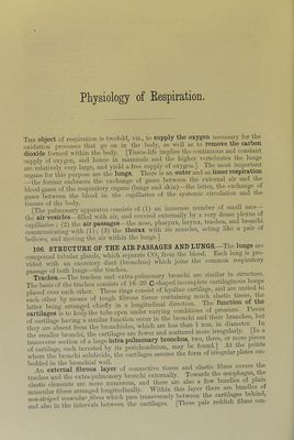
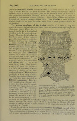
![stitute the trachealis muscle, and are attached to the inner surfaces of tlie carti- lages at a little distance from their free ends. The arrangement varies in different animals—thus, in the cat, dog, rahhit, and rat the muscular libres are attached to the external surfaces of the cartilages, while in the pig, sheep, and ox they are attached to their internal surfaces {Stirling).'\ Some muscular fibres are arranged longitudinally external to the transverse libres. The function of these muscular fibres is to irrevent too great distention when there is great pressure within the air-passages. The mucous membrane of the trachea consists of a basis of very fine connective-tissue, containing much adenoid tissue with numerous lymj)h-corpuscles. Xumerous elastic fibres are ar- ranged chiefly in a longitudinal direction under the basement mem- brane. They are also abundant in the deep layers of the posterior part of the membrane opposite the intervals between the cartilages. A small quantity of loose sub- mucous connective tissue contain- ing the large blood-vessels, glands, and lymphatics unites the mucous membrane to the perichondrium of the cartilages. The epithelium consists of a layer of columnar ciliated cells with several layers of immature cells under them. [The siqrerficial layer of cells is columnar and ciliated (fig. 147, h), while those lying imder them present a variety of forms, and below all is a layer of somewhat flattened squames, c, resting on the base- ment membrane, d. These squames constitute a layer quite distinct from the basement membrane, and they form the layer described as Debove’s membrane. They are active germinating cells, and play a most important part in connec- tion with the regeneration of the epithelium, after the superficial layers have been shed, in such conditions as bronchitis. Not un- frequently a little viscid mucus (a) lies on the free ends of the cilia. rraiisverse section of part of a human bronchus ( x 450). «, precipitated mueus ; h, ciliated columnar epithe- 1mm ; c, deep germinal layer of cells (Debove’s mem- brane); elastic basement membrane ; c, elastic fibres divided transversely (inner fibrous layer) ; f, bionchial muscle ; g, outer fibrous layer with leuco- cytes and pigment granules (black) below x >iass In tlie intermediate layer, the cells ° tissue. are more or less pyriform or battledorc-.shaped, with their long taimrim^ i>rocess inserted amongst the deeper layers of cells. According to Drasch, this lom> l)rocess is attached to one of these cells and is an outgrowth from it,’the xvliole constituting a “ foot-cell.”] Under tlie cpitlielium is tlie homogeneous basement membrane, throimh which fine canals pa.ss, connecting tlie cement of the epithelium with spaces in tlie mucosa, [flu.s membrane is well marked in the human trachea, where it plays an important part m many patliological condition,s, e.g., lironchitis. It is stained bright red with](https://iiif.wellcomecollection.org/image/b21981516_0001_0221.jp2/full/800%2C/0/default.jpg)
 No text description is available for this image
 No text description is available for this image
 No text description is available for this image