Volume 1
A text-book of human physiology : including histology and microscopical anatomy : with special reference to the requirements of practical medicine / by L. Landois ; translated from the seventh German edition, with additions, by William Stirling.
- Date:
- 1891
Licence: Public Domain Mark
Credit: A text-book of human physiology : including histology and microscopical anatomy : with special reference to the requirements of practical medicine / by L. Landois ; translated from the seventh German edition, with additions, by William Stirling. Source: Wellcome Collection.
Provider: This material has been provided by the Royal College of Physicians of Edinburgh. The original may be consulted at the Royal College of Physicians of Edinburgh.
99/602 page 59
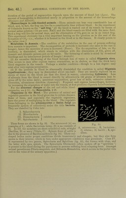
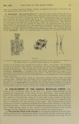
![exist ill the liumaii mesentery (Schohl). Similnr arrangenieiits may exist in connection with veins, giving rise to venous velict niivcibilici. 43. THE HEART. The muscular fibres of the mammalian heart consist of short (50 to 70 ^ ill man) very tine, transversely striated fibres, which are actual unieelliilar elements, devoid of a sarcolemma (15 to 25 ix. broad), and usually divided at their blunt ends, by which means they anastomose and form a network (fig. 34, A, B). The individual muscle-cells contain in their centre an oval nucleus, and are held together by a cement-substance, whicli is blackened by silver nitrate, and dissolved by a 33 per cent, solution of caustic potash. _ This cement is also dissolved by a 40 per cent, solution of nitric acid. The transverse striie are not very distinct, and not unfrequently there is an appearance of longitudinal striation, produced by a number of very small granules arranged in rows within the fibres. The fibres are gathered lengthwise in bundles, or fasciculi, surrounded and separated from each other by delicate processes of the perimysium. When the connective-tissue is dissolved by ])rolonged boiling, these Fig. 34. A, muscular fibres from the heart of a mammal, and C from a frog; B, transverse section of the cardiac fibres ; 6, connectiveTissue corpuscles ; c, capillaries. bundles can be isolated, and constitute the so-called “fibres” of the heart. The transverse sections of the bundles in the auricles are polygonal or rounded, while in the ventricles they are somewhat flattened. [The muscular mass of the heart is called the myocardium, and is invested by fibrous tissue. It is important to notice that the connective-tissue of the viscer.al pericardium (epicardium) is continuous with that of the endocardium by means of the perimysium suirounding the bundles of muscular fibres.] The fine spaces which exist between these bundles form narrow lacuna?, lined with epithelium, and constituting part of the lymphatic system of the heart. [The cardiac muscular fibres occupy an intermediate position between striped and plain muscular fibres. Although they are striped, they are involuntary, not being directly under the influence of the will, while they contract more, slowly than a voluntary muscle of the skeleton.] In the frog’s heart the muscular fibres are in shape elongated spindles, or fusiform, in this respect resembling the plain muscle-cells, but they are transversely striped (fig. 34, C). They are ea.sily isolated by means of a 33 per cent, solution of potash or dilute alcohol. 44. ARRANGEMENT OF THE CARDIAC MUSCULAR FIBRES.—The .study of the embryonic heart is the key to a proper understanding of tlie coni])licated arrangement of the fibres in the adult heart. The simple tubular lieart of the embryo has an outer circular and an inner longitudinal layer of filires. Tlie septum is formed later; hence, it is clear that a ]iart, at least, of the fibres must be common to the two auricles, and a part also to the two A’cntricles, since there is, originally, but one chamber in the heart. The muscular filnes of the atiricles are, however, completely separated, from those of the ventricles by the fibro- cartilaginous rings. In the auricles the fundamental arrangement of the embryonic fibres ])artly remains, while in tire ventricles it becomes obscured as the cavities underg(; a sac-like dilatation, and also become twisted in a s])iral manner. (1) The muscular fibres in the auricles are completely separated from the fibres of the ventricles by tlie fibrous rings which surround the auriculo-ventricular orifices, and which serve as aii attachment for tlie auriculo-ventricular valves (fig. 35, 1.).](https://iiif.wellcomecollection.org/image/b21981516_0001_0099.jp2/full/800%2C/0/default.jpg)


 No text description is available for this image
 No text description is available for this image
 No text description is available for this image