The surgical treatment of brain abcess by exposure and enucleation / Robert A. Groff and Francis C. Grant.
- Groff, Robert A. (Robert Armand), 1903-
- Date:
- [1940?]
Licence: In copyright
Credit: The surgical treatment of brain abcess by exposure and enucleation / Robert A. Groff and Francis C. Grant. Source: Wellcome Collection.
5/34 page 929
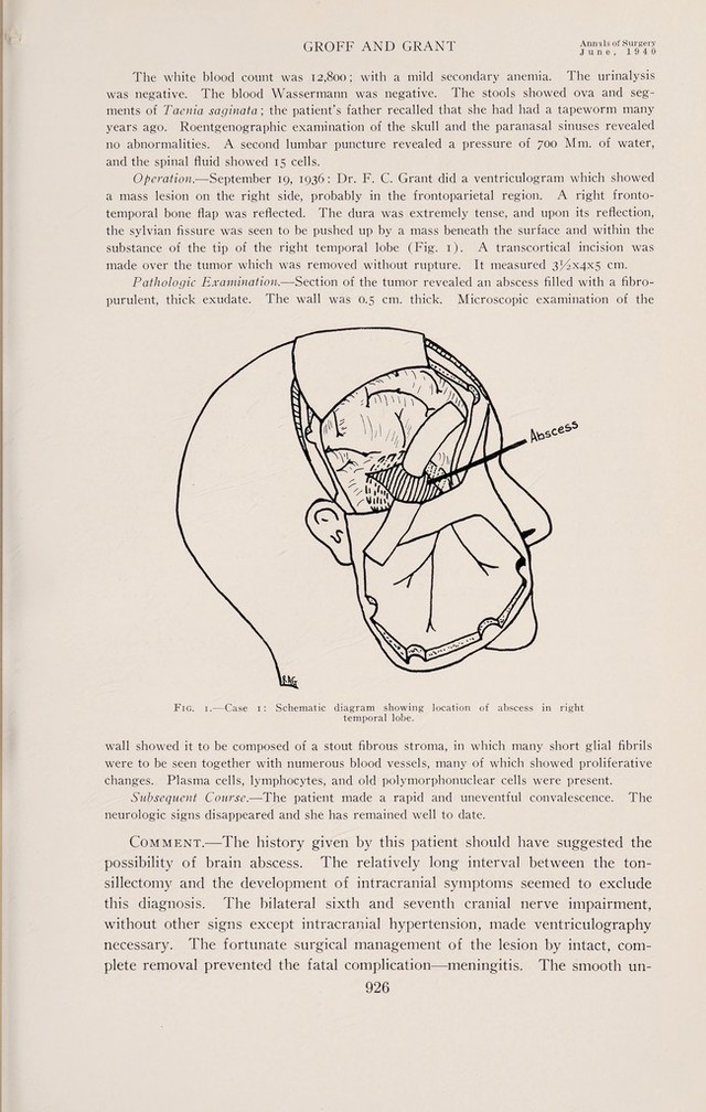
![Number 6 A complicated convalescence and the continuance of the patient s good health up to the present time, two and one-half years, commend this method of treatment. Case 2.—Synopsis: Symptoms for 14 weeks. Signs suggestive of expanding left cerebral lesion. Cannula struck abscess during ventriculography. Abscess exposed and removed without rupture. Recovery. J. F. M., Hosp. No. 35238, male, age 26, was admitted to the University Hospital, January 12, 1937, having been referred to Dr. F. C. Grant by Dr. Marshall W. Dyer, Syracuse, N. Y. The patient was well until 14 weeks before admission to the hospital, when he began complaining of projectile vomiting. This vomiting bore no relationship to meals or time of day and continued up until the time of admission. One week after the onset he developed left-sided headache which subsequently became generalized. Shortly afterward he had periodic attacks of dimness of vision. While walking he noticed he tended to deviate to the right and on numerous occasions became dizzy when bending ovei. jrIG> 2.—Case 2: Schematic diagram showing location of abscess in left occipital lobe. Physical Examination,—The patient was normal except for dental and tonsillar sepsis. Neurologic examination revealed a fair mental orientation, dysarthesia with test phrases, left sixth nerve palsy, choked disks of four to five diopters, positive tremnor sign on the right, abortive ankle clonus on the right together with mild inciease in reflexes on the right side of the body, dysnergia in finger to nose test on both sides, unsteady gait, and poor associated movements on the right side. Temperature, pulse and respirations were normal. The leukocyte count 10,600, and the Wassermann negative. The visual fields showed a marked contraction of both temporal fields, especially on the right, and a com¬ plete right homonomous hemianopia. Roentgenograms of the skull showed erosion of the posterior clinoids and dorsum of the sella with some forward displacement of the top of the dorsum. Operation,—]anuary 22, 1937: Dr. F. C. Grant performed a ventriculogram for the purpose of localization. A solid mass was encountered by the cannula when attempting to enter the posterior horn of the left lateral ventricle. Predicated upon this, a left](https://iiif.wellcomecollection.org/image/b30631592_0005.jp2/full/800%2C/0/default.jpg)