The surgical treatment of brain abcess by exposure and enucleation / Robert A. Groff and Francis C. Grant.
- Groff, Robert A. (Robert Armand), 1903-
- Date:
- [1940?]
Licence: In copyright
Credit: The surgical treatment of brain abcess by exposure and enucleation / Robert A. Groff and Francis C. Grant. Source: Wellcome Collection.
6/34 page 930
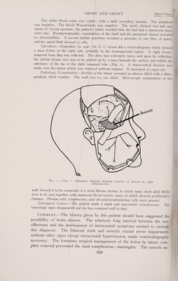
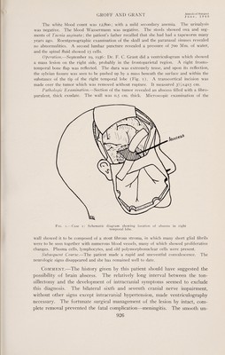
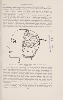
![GROFF AND GRANT Annals cl Surgery June, 1940 occipitoparietal bone flap was reflected. On the cortex, in the occipital lobe (Fig. 2), a grayish-yellow, massive tumor was exposed. This tumor was carefully dissected from its bed without damaging it. After complete hemostasis had been secured, the wound was closed. The tumor measured approximately 6x4x4 cm. Pathologic Examination, Section of the tumor revealed it to be an abscess containing thick greenish pus. The wall was quite thick and contained a few large blood vessels Microscopic examination of the wall showed it to be composed of a dense fibrous struc¬ ture arranged in parallel rows. Among these fibers were numerous fibroblasts and scat¬ tered polynuclear cells. The patient made a rapid and uneventful convalescence. On discharge practically all of the neurologic signs had disappeared but the disks showed a choking of two diopters. Subsequent Course.—The patient has been seen since operation. The vision and visual fields have improved. However, in the six months following removal of the abscess three generalized major convulsions have occurred. He was given small amounts of phenobarbital and has had no attacks for the past 18 months. Comment.-—This patient presented essentially the same problem as the case just described. The history was longer and the neurologic signs were suggestive but not conclusive of exact localization. The abscess was exposed by an osteoplastic flap and completely removed without damage. The rapid convalescence and the necessity of only 20 days’ hospitalization further empha¬ size the rationale of this method of treatment. CaSe 3'~Synops{s: ^acranial symptoms for two weeks following mild head injury and streptococcic sore throat. Signs of intracranial hypertension and. a right parietal cswn. Craniotomy, tap of abscess by exploring cannula, and complete enucleation. Re¬ covery. S. K., Hosp. No. 39361, male, age 21, was admitted to the University Hospital June 6 1938, having been referred by Dr. C. C. Neff, York, Pa. The patient had been perfectly well until two weeks before admission, when he bumped his head on a beam. He did not become unconscious nor were there any ill effects from this accident. Shortly afterward the patient developed a sore throat which lasted one week. The infection was alleged to be caused by the streptococcus. One week before admission he had an attack in which both arms and legs “stiffened,” but no loss of consciousness occurred. This attack began m.the left arm and sPread to involve the rest of the body. It lasted approximately ten minutes. During the next three days he had three to four similar attacks. Since then tie patient has become progressively more dizzy, especially when attempting to walk. At about the same time these attacks began he developed right frontal headaches. For the several days before admission the left arm and hand had become weak. Physical Examination.—The patient was acutely ill. Temperature 99° F., pulse 80, respirations 20. Neurologic examination : The patient was definitely lethargic, the eye- grounds showed a bilateral papilledema of between two to three diopters. The visual e ds, to gross tests, were normal. There was a left central facial weakness. The corneal reflex was decreased on the left. The left arm and hand were weak in all movements and a similar but less marked weakness was demonstrated in the left leg. Reflexes in the left arm were increased over those in the right. In the lower extremities, the reflexes were l aterally exaggerated but equal. A sustained ankle clonus was present on both sides. 6 1fft1S1,dei of the body’ mcluding the face, showed a reduction to all forms of sensation, and the left hand showed a loss of stereognostic sense. White blood count 20,300- spinal fluid pressure was 300 Mm. of water, and showed four cells per cubic millimeter Operation.-]une 2, 1938: Dr. L. Weinberger performed bilateral frontal and parietal rephmes. No hematoma was found. The ventricles were tapped and the left was found to be larger than the right. On June 3, 1938, Dr. F. C. Grant reflected a right frontoparietal bone flap. The](https://iiif.wellcomecollection.org/image/b30631592_0006.jp2/full/800%2C/0/default.jpg)