Anatomy and physiology : designed for academies and families / by Calvin Cutter.
- Calvin Cutter
- Date:
- 1847
Licence: Public Domain Mark
Credit: Anatomy and physiology : designed for academies and families / by Calvin Cutter. Source: Wellcome Collection.
Provider: This material has been provided by the National Library of Medicine (U.S.), through the Medical Heritage Library. The original may be consulted at the National Library of Medicine (U.S.)
89/352
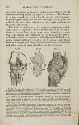
![Fig- 41. Fig. 12. Fig. 41. The ligaments of the pelvis and hip joint. 1, The lower part of the anterior dgament of the vertebrae. 2, A ligament which extends from one of the vertebra to the sacrum. 3, A ligament which extends from the vertebrae to the ilium. 4, Liga- ments that connect the ilium and sacrum. 6, The obturator membrane. 6, Poupart's ligament. 7, Gimbernat's ligament. 8, The capsular ligament of the hip joint. 9, The ilio-femoral or accessary ligament. Fig. 42. 1, The sacro-iliac ligament. 2, The acetabulum. 5. The head of the thigh bone. 4, The neck of the thigh bone. 3, The ligament that binds the head of the thigh bene to the bottom of the acetabulum. Fig. 43. Fig. 44. Fig. 43. The anterior ligaments of the knee joint. 1, The tendon of the quadriceps extensor muscle of the leg. 2, The patella. 3, The anterior ligament of the patella, near its insertion. 4,4, The synovial membrane. 5, The internal lateral ligament. 6. The long external lateral ligament. 7. The anterior and superior ligament that unites the fibula to the tibia. Fig. 44. The posterior ligaments of the knee joint. ], The ligament of Wlnslow. 2 The tendon of seml-membianosus muscle. 3, Its insertion, showing the expansion of Its fibres. 5, The internal lateral ligament. 6, 7, The external lateral ligament. 8, The tendon of a muscle cut off. t>, The post'dor tibial ligament.](https://iiif.wellcomecollection.org/image/b21113002_0089.jp2/full/800%2C/0/default.jpg)