The Erasmus Wilson lectures on the anatomy and pathology of the eye : delivered at the Royal College of Surgeons on Feb. 12th, 14th, and 16th, 1900 / by E. Treacher Collins.
- Collins, E. Treacher (Edward Treacher), 1862-1937.
- Date:
- [1900]
Licence: In copyright
Credit: The Erasmus Wilson lectures on the anatomy and pathology of the eye : delivered at the Royal College of Surgeons on Feb. 12th, 14th, and 16th, 1900 / by E. Treacher Collins. Source: Wellcome Collection.
Provider: This material has been provided by UCL Library Services. The original may be consulted at UCL (University College London)
39/602 (page 7)
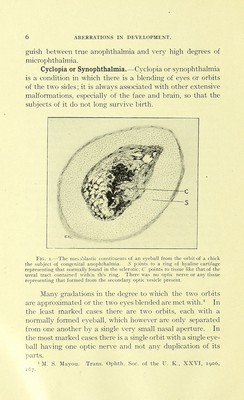
![The fusion of the two orbits results in tiie fronto-nasal process, from which the nose should have developed being pushed forward and formed into a proboscis which hangs down from above the orbit (Fig. 2). When one central orbit is formed it is surrounded by four eyelids, the palpebral aperture between whicii is roughly diamond-shaped (Figs. 3, 4). The upper canal- iculi become suppressed; the lower are present but termi- nate in a blind canal. Fig. 2.—Human cyclops with rxtu nic ili L!;rcc ol iiiiiroijhthalmia. Note proboscis with opening at the end (the upper hole is due to a pin marlt. The degree of fusion of the two eyes is very \'arious; often besides the changes resulting from the mere fusion there are other maldevelopments. The fused eyes may be microphthalmic or abnormally large. When one eye is present without any duplication of its stmctures, the cornea is generallv of an o\'al sha]ie with the long axis horizontal. All the structures in the eye may be single and the lenses alone show signs of fusion. The cornea and sclerotic mav be single, but the iris, choroid, retina, lens and vitreotis](https://iiif.wellcomecollection.org/image/b21286292_0039.jp2/full/800%2C/0/default.jpg)