Holden's manual of the dissection of the human body / edited by Luther Holden and John Langton.
- Date:
- 1879
Licence: Public Domain Mark
Credit: Holden's manual of the dissection of the human body / edited by Luther Holden and John Langton. Source: Wellcome Collection.
647/712
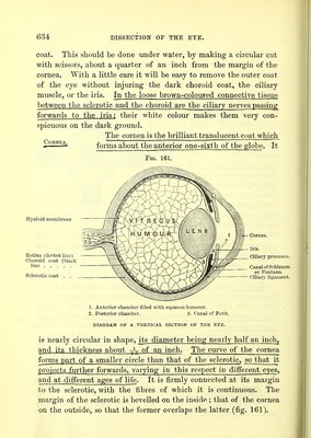
![Fig. 160. OF THE RECTI MUSCLES WITH .NTERIOR CILIARY ARTERIES. one-sixth being completed by the cornea. The thickest part of the sclerotic coat is at the back of the globe (fig. 161) ; the thinnest is a short distance behind the cornea.* The back of the sclerotic is perforated by the optic nerve, which enters it about one-tenth of an inch on the nasal side of the axis of vision. The optic nerve at its entrance into the sclerotic is .much constricted, and instead of passing through a single aperture in this coat, enters it through .a porous network of fibrous tissue, called the lamina cribvosa.] The sheath of the optic nerve becomes continuous with the sclerotic where it perforates this coat. Around the optic nerve the sclerotic is perforated by the ciliary arteries, veins, and nerves,INSEjmoN for the supply of the choroid and iris. About a quarter of an inch from the cornea the sclerotic receives the insertion of the recti muscles ; here also it transmits. the anterior ciliary arteries, which ran forward along the tendons of these muscles, and form a vascular ring around the margin of the cornea (fig. 160). The sclerotic is composed of connective tissue arranged in bundles, which run, some longitudinally, some transversely. The longitudinal fibres are the most -external and abundant. Under the microscope numerous connective-tissue corpuscles may be seen rilling cell-spaces, similar to those found-in the cornea, but not so abundant, and containing pigment-granules. Between these may be demonstrated fine elastic fibres. The inner surface of the sclerotic is coated with a thin layer of connective tissue, lamina fusca, in which are found some pigment-cells. To examine the cornea, it should be removed with the sclerotic * The greatest thickness posteriorly is about the ^th of an inch; its least thick- ness infront is about ~th of an inch. f_ In the centre of the lamina cribrosa is an opening larger than the rest, Tvhich transmits the arteria centralis_retins8.](https://iiif.wellcomecollection.org/image/b20417780_0647.jp2/full/800%2C/0/default.jpg)