Holden's manual of the dissection of the human body / edited by Luther Holden and John Langton.
- Date:
- 1879
Licence: Public Domain Mark
Credit: Holden's manual of the dissection of the human body / edited by Luther Holden and John Langton. Source: Wellcome Collection.
675/712
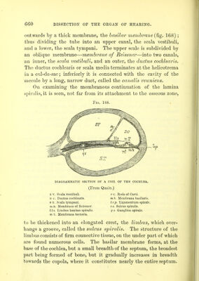
![It consists of a firm, fibrillated tissue, which is probably formed, ni any rate on its upper surface, of a structure closely resembling the organ of Corti. The membrane which separates the scala vestibuli and the * ductus cochlearis is a delicate almost structureless layer, the mt m- brane of Reissner. It appears to be composed of connective tissue, lined on its vestibular surface with flattened connective-tissiu cells, and on its cochlear surface with squamous epithelium. At the point of attachment of the basilar membrane with the outer wall of the cochlea may be seen a triangular projection, which, formerly described as a muscle, (cochlearis muscle), is now generally believed to be a collection of connective-tissue cells, and called the ligamentum spirale. Organ of The organ of Corti is a highly complex structure, Cohti. placed on the upper surface of the basilar mem- brane, and the floor of the ductus cochlearis. The central part of the organ of Corti is formed by two sets of slanting rods—inner and outer rods of Corti* which rest against each other at their upper extremities, thus forming a triangular tunnel beneath them, filled in the recent state with endolymph. On the inner side of the inner rods is a single row of cells tipped with ciliated processes, called the inner hair-cells ; and on the outer side of the outer rods are three rows of similar cells, termed the outer hair-cells. The only remaining membrane to be described is the tectorial membrane, which lies above and parallel to the basilar membrane, but does not extend much more than half-way over it. Il is con- nected on its inner side with the limbus spiralis, and is then con- tinued outwards, overlying and resting upon the rods of Corti, and ends in a free extremity. It is a strong, elastic membrane, distinctly fibrous, especially upon its inner and thicker part. The cochlear division of the auditory nerve (the vestibular lias already been described,p. 659) is a short, thick branch, which breaks up into numerous filaments at the bottom of the meatus auditurius * The inner rods are stated to be more numerous than the outer, in the pro] ortiuu of 6,000 of the inner to 4,500 of the outer rods.](https://iiif.wellcomecollection.org/image/b20417780_0675.jp2/full/800%2C/0/default.jpg)