The liver, spleen, pancreas, peritoneal relations, and biliary system in monotremes and marsupials / by William Colin MacKenzie.
- William Colin Mackenzie
- Date:
- 1918
Licence: In copyright
Credit: The liver, spleen, pancreas, peritoneal relations, and biliary system in monotremes and marsupials / by William Colin MacKenzie. Source: Wellcome Collection.
23/184 page 3
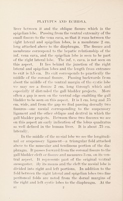
![exit of the vena cava, meet the peritoneal folds connect- ing liver to diaphragm. From this point also where it is continuous with the suspensory ligament we see a mem- brane attached above to the diaphragm and curving down on the inner side of the left lateral lobe. At the dorsal curved portion of the oblique fissure it is continuous with the gastro-liepatic omentum. In a large male Platy- pus the lateral (dextro-sinistral) measurement of the liver was 13 cm. and dorso-ventral measurement was 9 cm. Laterally, cystic was 10 cm., left lateral was 7 cm., right lateral including spigelian was 8 cm. (b) Echidna.—Here we distinguish the same divi- sions as in the Platypus, viz. mesial or cystic in front sep- arated into a large right and smaller left portions by the suspensory ligament, and dorsally the right and left lat- eral lobes separated from the former by the coronal fis- r i/ sure. The cystic lobes form less of the anterior margin of the liver than in Platypus. The suspensory ligament runs obliquely backwards and to the right (not mesially) from the ventral border to the coronal fissure (inf. v. cava.). Its length is 1.5 cm. and it is attached above to the mus- cular and tendinous part of the diaphragm. There is no gall bladder fissure as in the Platypus, i.e. the gall bladder does not project on the diaphragmatic surface. In only one of a series of eight specimens was there a thinning of the liver substance over the gall bladder with a slight indentation on the ventral margin. This was to the right of the suspensory ligament. Though the coronal fissure may completely separate the cystic from the lateral lobes yet we frequently see evidence of fusion dorsally between the right lateral and right cystic and left lateral with the left cystic lobes, and especially the latter. Thus we have a right and left coronal fissure and the left corona] mav be 1.25 cm. from the inf. v. cava. There 9 is a more complete differentiation of the spigelian from](https://iiif.wellcomecollection.org/image/b29821125_0023.jp2/full/800%2C/0/default.jpg)