A manual of physiology : for the use of junior students of medicine / by Gerald F. Yeo.
- Date:
- 1884
Licence: Public Domain Mark
Credit: A manual of physiology : for the use of junior students of medicine / by Gerald F. Yeo. Source: Wellcome Collection.
Provider: This material has been provided by the Royal College of Physicians of Edinburgh. The original may be consulted at the Royal College of Physicians of Edinburgh.
620/676 page 596
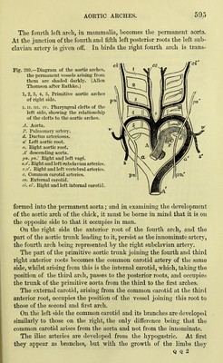
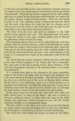
![become so much larger tliat after birth they appear to be the main branches from the point of division of the aorta, the hypogastric arteries now being merely small branches of the iliac vessels. With the development of the organs and limbs, vessels in connec- tion with those above described arise in the mesoblast. It is, how- ever, beyond the scope of this work to describe in detail the origin ©f the lesser vessels. Fig. 290. A. Plan of principal veins of the foetus of about four weeks old. 7>. Veins of the liver at an earlier period. C. Veins after the establishment of the placental circulation. D. Veias of the liver at the same period. ./. Primitive jugular veins. dc. Ducts of Cuvier. ca. Cardinal veins. ci. Inferior vena cava. I. Ductus venosus. n. Umbilical vein. p. Portal vein. o. Vitelline vein. <^r. External iliac veins. o'. Right vitelline vein. .?<'. Right umbilical vein. Hepatic veins (vense reve- hentes). ])'p'. Yenve advehentes. m. Mesenteric veins. az. Azygos vein. ca'. Remains of left cardinal vein. Subclavian vein. //. Cross branch from left jugu- lar which becomes the left brachio-cephalic veia. ri. Right innominate vein. s.s. Subclavian veins. ?t. Hypogasti'ic veins. i/. Division of inferior vena cava into the common iliac veins. Venous System — The blood is returned from the head by the iwo primitive jugulars, v, hich unite with the ^irdinal veins conveying](https://iiif.wellcomecollection.org/image/b21932943_0620.jp2/full/800%2C/0/default.jpg)