A manual of physiology : for the use of junior students of medicine / by Gerald F. Yeo.
- Date:
- 1884
Licence: Public Domain Mark
Credit: A manual of physiology : for the use of junior students of medicine / by Gerald F. Yeo. Source: Wellcome Collection.
Provider: This material has been provided by the Royal College of Physicians of Edinburgh. The original may be consulted at the Royal College of Physicians of Edinburgh.
631/676 page 607
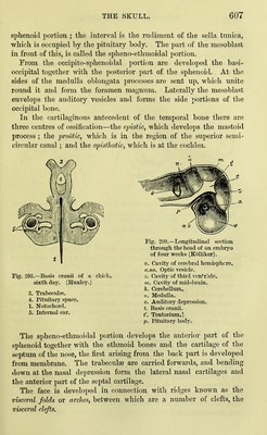
![sphenoid portion ; the interval is the rudiment of the sella tunica, which is occupied by the pituitary body. The part of the mesoblast in front of this, is called the spheno-ethmoidal portion. From the occipito-sphenoidal portion are developed the basi- occipital together with the posterior part of the sphenoid. At the sides of the medulla o1)longata processes are sent up, which unite round it and form the foramen magnum. Laterally the mesoblast enveloj)s the auditory vesicles and forms the side portions of the occipital bone. In tlie cartilaginous antecedent of the temporal bone there are three centres of ossification—the einotic, which develops the mastoid process ; the i^roUtic, which is in the region of the superior semi- circular canal ; and the opisthotic, which is at the cochlea. Fig. 298.—Basis cranii of a chick, sixth day. (Huxley.) 3. Trabeculee. 4. Pituitary space. 1. Notochord. 5. Internal ear. Fig. 299.—Longitudinal section through the head of an embryo of four weeks (Kolliker). V. Cavity of cerebral hemisphere. a.no. Optic vesicle. z. Cavity of third ventricle. m. Cavity of mid-brain. h. Cerebellum. n. Medulla. 0. Auditory depression. t. Basis cranii. t'. Tentorium.] p. Pituitary body. The spheno-ethmoidal portion develops the anterior part of the sphenoid together with the ethmoid bones and the cartilage of the septum of the nose, the first arising from the back part is developed from membrane. The trabeculae are carried forwards, and bending down at the nasal depression form the lateral nasal cartilages and the anterior part of the septal cartilage. The face is developed in connection with ridges known as the visceral folds or arches, between which are a number of clefts, the visceral clefts.](https://iiif.wellcomecollection.org/image/b21932943_0631.jp2/full/800%2C/0/default.jpg)