The evolution of man : a popular scientific study / by Ernst Haeckel. Translated from the 5th (enl.) ed. by Joseph McCabe.
- Ernst Haeckel
- Date:
- 1905
Licence: In copyright
Credit: The evolution of man : a popular scientific study / by Ernst Haeckel. Translated from the 5th (enl.) ed. by Joseph McCabe. Source: Wellcome Collection.
Provider: This material has been provided by The University of Leeds Library. The original may be consulted at The University of Leeds Library.
333/392 (page 307)
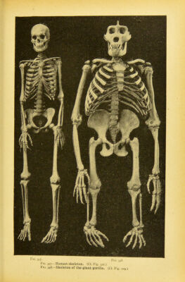
![parts, tlie lowest living races of inea (the Veddahs of Ceylon, Fig. 344) are midway between the chimpanzee (Fig. 343) and the European (Fig. 345). More consider- able are the differences in structure and the proportions of the various parts be- tween the different genera of anthropoid apes (Figs. 278-2S2); and still greater is the morphological distance between these and the lowest apes (the Cynopitheca). Here,again, impartial and thorough ana- tomic comparison confirms the accuracy of Huxley's pithecometra principle (p. 171). The complete unity of structure which is thus revealed by the comparative anatomy of the limbs is fully confirmed bv their embryology. However different the extremities of the four-footed Craniotes may be in their adult state, they all de- velop from the same rudimentary struc- ture. In every case the first trace of the limb in the embryo is a very simple pro- tuberance that grows out of the side of the hyposoma. These simple structures develop directly into fins in the fishes and Dipneusts by differentiation of their cells. In the higher classes of Vertebrates each of the four takes the shape in its further growth of a leaf with a stalk, the inner half becoming narrower and thicker and the outer half broader and thinner. The inner half (the stalk of the leaf) then divides into two sections—the upper and lower parts of the limb. Afterwards four shallow indentations are formed at the free edge of the leaf, and gradually deepen ; these are the intervals between the five toes (Fig. 174). The toes soon mtike their appearance. But at first all five toes, both of fore and hind feet, are connected by a thin membrane like a swimming-web ; they remind us of the original shaping of the foot as a paddling fin. The further development of the limbs from this rudimentary structure takes place in the same way in all the Vertebrates according to the laws of heredit}-. The embryonic development of the muscles, or active organs of locomotion, is not less interesting than that of the skeleton, or passive organs. But the comparative anatomy and ontogeny of the muscular system are much more dillli- cult and inaccessible, and consequently have hitherto been less studied. We can therefore only draw some general phylo- genetic conclusions therefrom. It is incontestable that the musculature of the Vertebrates has been evolved from that of lower Invertebrates ; and among these we have to consider especially the unarticulated Vermalia. They have a simple cutaneous muscular layer, develop- ing from the mesoderm. This was after- wards replaced by a pair of internal lateral muscles, that developed from the middle wall of the ccrlom-pouches ; we still find the first rudiments of the muscles arising from the muscle-plate of these in the embryos of all the Vertebrates (of. Figs. 124, 158-60, 222-4 I'l t'le unarticu- lated stem-forms of the Chordonia, which we have called the Prochordonia, the two coelom-pouches, and therefore also the muscle-plates of their walls, were not yet segmented. A great advance was made in the articulation of them, as we have followed it step by step in the Amphioxus (Figs. 124, 158). This segmentation of the muscles was the momentous historical process with which vertebration, and the development of the vertebrate stem, began. The articulation of the skeleton came after this segmentation of the muscular system, and the two entered into very close corre- lation. The episomites ordorsal coelom-pouches of the Acrania, Cyclostomes, and Selachii (Fig. 161 h) first develop from their inner or median wall (from the cell-layer that lies directly on the skeletal plate [sk] and the medullary tube [pir]) a strong muscle- plate (mpJ. By dorsal growth (wJ it also reaches the external wall of the coelom-pouches, and proceeds from the dorsal to the ventral wall. From these segmental muscle-plates, which are chiefly concerned in the segmentation of the Vertebi-ates, proceed the lateral muscles of the stem, as we find in the simplest form in the Amphioxus (Fig. 210); By the formation of a horizontal frontal septum they divide on each side into an upper and lower series of myotomes, dorsal and ventral lateral muscles. This is seen with typical regularity in the transverse section of the tail of a fish (Fig. 346). From these earlier lateral muscles of the trunk develop the greater part of the subsequent muscles of the trunk, and also the much later muscular buds of the limbs.' ' The ontog-cny of the imiscles is mostly cenogrenetic. The greater part of the nniscles of the' head (or the visceral musclos) beloiij;: oriffinally lo tlie hyposoma of the vertebrate organism, and develop from'the wall of the liyposomites or ventral ca;loni-pouches. This also applies originally to the prini.try muscles of the limlis, as these too belong phylogenclically to the liyposoma. (Ct. Chapter XIV.)](https://iiif.wellcomecollection.org/image/b21508616_0333.jp2/full/800%2C/0/default.jpg)