Treatise on the diseases of the eye : including the anatomy of the organ / by Carl Stellwag (von Carion) ; translated from the fourth German edition and edited by D.B. St. John Roosa, Charles S. Bull, and Charles E. Hackley.
- Karl Stellwag von Carion
- Date:
- 1873
Licence: Public Domain Mark
Credit: Treatise on the diseases of the eye : including the anatomy of the organ / by Carl Stellwag (von Carion) ; translated from the fourth German edition and edited by D.B. St. John Roosa, Charles S. Bull, and Charles E. Hackley. Source: Wellcome Collection.
Provider: This material has been provided by the Harvey Cushing/John Hay Whitney Medical Library at Yale University, through the Medical Heritage Library. The original may be consulted at the Harvey Cushing/John Hay Whitney Medical Library at Yale University.
83/954 (page 53)
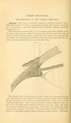
![1. The greater number or all of the wandering cells, or those newly formed by proliferation, often undergo fatty degeneration, together with the inter-cellular sub- stance separated from them, which contains more or less fibrin; ]ms is formed in the true tissue of the cornea, which being soon involved, becomes opaque, soft, and is finally also destroyed, becoming a mass of fatty detritus. From the surface of the cornea such products soon fall, as the sub-epithelial layer is rapidly destroyed. The result is a more or less extensive ulcer. But within the cornea pus is retained for some time, where, so long as it is inclosed by corneal substance, it is called an ahseess. The laminated structure of the cornea, then, not unfrequently allows a por- tion of the fluid-pus to pass between the lamella? and collect in quite large quantities in the inter-lamellar spaces, near the corneal border, and thus an onyx is formed. 2. Just as often the inflammatory products go on to a higher degree of develop- ment. The collection of formative cells which lie superficially are especially inclined to this. These are quickly divided into two layers, which are connected to each other by variously shaped processes, and are usually sharply defined from each other. The elements of the anterior layer become epithelial cells, while those of the posterior layer grow, and by a change into the fusiform shape as well as by a grad- ual formation of an intercellular substance which soon becomes striated, reminds us of connective tissue or true corneal substance. In this granulation layer vessels soon become evident, which proceed from those in the limbus conjunctivalis, and permeate the layer of neoplastic cells, ahvays running towards the center, and finally form a net-work of large trunks. These are then continued in greatly dila- ted conjunctival veins. It seems as if the blood at first ran in sharply bounded fissures between the elements of the granulation layer, for actual walls are not seen at an early period (Jwanoff). They then appear as branched tubes, which are closely covered by fusiform cells {His, Niemet&chek) which under some circumstances may become a very dense adventitious membrane. The greater number and often even all of the neoplastic vessels run above Bowman's membrane, where this still exists. This membrane is frequently destroyed at an early period, however, at least some parts of it. Thus there ceases to be any boundary membrane for the vessels that run through the whole granular layer. The blood, which runs in vessels visible to the naked eye, must be considered as venous, in consonance with the nature of the emergent conjunctival trunks, although its color is a light red. This color is explained by the superficial situation of the vessels, which allows the oxidizing influence of the atmospheric air. This appearance of blood-vessels is so marked, that for a long time it has been considered the indication of a peculiar form of corneal inflammation, vascular ler- atitis. The changes in the granulation layer which have been described are apt to increase with the duration of the disease. They are found most advanced, and are therefore most distinctly seen when a chronic inflammation has existed for a long time ; especially when the irritation has been slight, and the condition has thus the character of pannus. Then the granulation layer is developed into true connective tissue, in which are found quite large vessels with dense adventitious tissue. Newly formed elements shut up in the true corneal tissue show, generally speaking, a much less tendency to higher development and the formation of blood-vessels, except When the layers affected have approached the surface by ulceration or traumatic less of substance of the layers over them. Then true corneal substance usually develops from these newly-formed elements, which more or less completely fills the existing](https://iiif.wellcomecollection.org/image/b21002319_0083.jp2/full/800%2C/0/default.jpg)