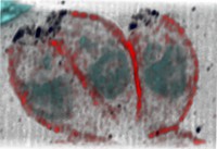
Structured-illumination micrograph (SIM) of three tachyzoites, a developmental stage in the Toxoplasma gondii parasite life cycle. The plasma membrane (red), an apical protein (black) and DNA (blue) is visible in each. Toxoplasma gondii is a parasitic protozoa that causes toxoplasmosis. Toxoplasmosis is a common infection in birds and mammals. If infected during pregnancy (for example from eating raw or undercooked meat or from contact with cats), the mother can pass the infection onto her unborn child (congenital toxoplasmosis). This can result in serious health complications for the child. Each parasite measures 10 micrometres. Structured-illumination microscopy (SIM) is one type of super-resolution microscopy. Super-resolution microscopy 2015