Guide to the exhibition of specimens illustrating the modification of the structure of animals in relation to flight.
- National Museum of Ireland, Merrion Street
- Date:
- 1913
Licence: In copyright
Credit: Guide to the exhibition of specimens illustrating the modification of the structure of animals in relation to flight. Source: Wellcome Collection.
Provider: This material has been provided by The Royal College of Surgeons of England. The original may be consulted at The Royal College of Surgeons of England.
18/96 page 6
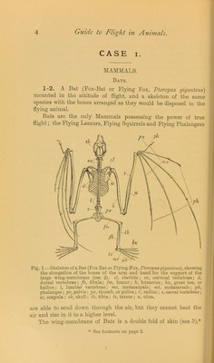
![the Bats of the family Emballonuridae the phalangeal portion of the third finger bends back on the dorsal surface of the wing, and to such an extent that the first phalangeal bone comes to lie parallel with the metacarpal bone. 5. Eight wing of a Bat {Pteropus vampyrus) with the mem- brane between the ulnar bone and the fifth digit partially split ^ to show that it is composed of two separable layers of skin. Compare with this the preparation of the Moth’s wing, 101. 6. Portion of skeleton of a Bat {Pteropus vampyrus'), left side, ] showing the extent of the keel of the sternum for the attachment of the depressor muscles of the wings (fig. 2). Fig. 2. —Thoracic skeleton of a large Fruit Bat {Pteropus vampyrus), together f with the cervical and lumbar Vertebrae, seen from the left side, k, the : keel of the sternum, to which the depressor muscles of the wings are attached (see 6). 7-8. Dissection of the shoulder of a Bat (Fruit Bat, Pteropus ] vampyrus) and explanatory sketch (see fig. 3). In Bats the principal muscles concerned in elevating and depressing the ; wing during flight are the dcltoideus and the pcctoralis major. ' The deltoideus (shown in blue in the explanatory sketch) on con- traction raises the wing; it is inserted into the upper * part of the first bone of the wfing (humerus), and takes its origin from the upper surface of the shoulder blade (scapula) and from a high- standing process of it (the acromion). The scapula itself is not a rigid part of the skeleton, but is held in position by the contraction of the trapezius and other muscles. The pcctoralis imjor (shown in red in the explanatory sketch) is the largest muscle in the body of the Bat, and by its contraction brings about the depression of the wing during flight. It arises mainly from the side of the * I.B. dorsal in the extended wing.](https://iiif.wellcomecollection.org/image/b22470888_0020.jp2/full/800%2C/0/default.jpg)