Principles of anatomy and physiology for physical training instructors in the Royal Air Force.
- Date:
- 1946
Licence: Public Domain Mark
Credit: Principles of anatomy and physiology for physical training instructors in the Royal Air Force. Source: Wellcome Collection.
49/194 page 39
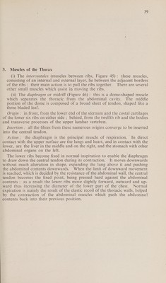
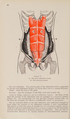
![3. Muscles of the Thorax (i) The intercostales (muscles between ribs, Figure 45): these muscles, consisting of an internal and external layer, lie between the adjacent borders of the ribs ; their main action is to pull the ribs together. There are several other small muscles which assist in moving the ribs. (11) The diaphragm or midriff (Figure 46): this is a dome-shaped muscle which separates the thoracic from the abdominal cavity. The middle portion of the dome is composed of a broad sheet of tendon, shaped like a three bladed leaf. Origin : in front, from the lower end of the sternum and the costal cartilages of the lower six ribs on either side ; behind, from the twelfth rib and the bodies and transverse processes of the upper lumbar vertebre. Insertion : all the fibres from these numerous origins converge to be inserted into the central tendon. Action: the diaphragm is the principal muscle of respiration. In direct contact with the upper surface are the lungs and heart, and in contact with the lower, are the liver in the middle and on the right, and the stomach with other abdominal organs on the left. The lower ribs become fixed in normal inspiration to enable the diaphragm to draw down the central tendon during its contraction. It moves downwards without much alteration in shape, expanding the lung above it and pushing the abdominal contents downwards. When the limit of downward movement is reached, which is decided by the resistance of the abdominal wall, the central tendon becomes the fixed point, being pressed hard against the abdominal contents ; as a result the lower ribs move slightly forward, outward and up- ward thus increasing the diameter of the lower part of the chest. Normal expiration is mainly the result of the elastic recoil of the thoracic walls, helped by the contraction of the abdominal muscles which push the abdominal] contents back into their previous position.](https://iiif.wellcomecollection.org/image/b32176867_0049.jp2/full/800%2C/0/default.jpg)

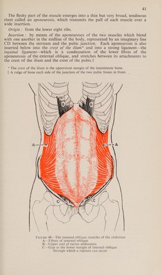
 No text description is available for this image
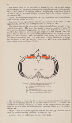
 No text description is available for this image
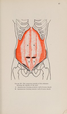
 No text description is available for this image