Volume 1
Contemporary classics in the life sciences / edited by James T. Barrett.
- Date:
- [1986-]
Licence: Attribution-NonCommercial 4.0 International (CC BY-NC 4.0)
Credit: Contemporary classics in the life sciences / edited by James T. Barrett. Source: Wellcome Collection.
33/400 page 11
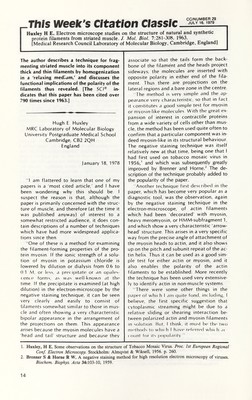
![CC/NUMBER 49 -This week's Citation Classic 0E 6 Anderson T F. Techniques for the preservation of three-dimensional structure in preparing specimens for the electron microscope. Trans. NY Acad. Sci. 13 :130 -4, 1951. [Johnson Foundation, University of Pennsylvania, Philadelphia, PA] Distortions of biological and other speci mens by surface tension are eliminated by first heating the ambient fluid above its critical point (where its surface tension vanishes) and then allowing the now gaseous fluid to escape. Thus prepared, delicate specimens retain their three-dimensional structures. [The SC/® indicates that this paper has been cited in over 1,060 publica tions since 1961.] Thomas F. Anderson Institute for Cancer Research Fox Chase Cancer Center Philadelphia, PA 19111 September 15, 1982 Originally, most aqueous specimens were simply dried in air for study in the high vacuum of the electron microscope. When this is done, the surface tension of the evaporating water flattens most objects on to the supporting membrane. Thus, our early studies designed to show whether tailed bacteriophages adsorb to their host bacteria by their heads or by their tails gave equivocal results It seemed to me that one could elimi nate surface tension forces by heating the ambient liquid to a temperature above its critical point where it changes impercep tibly into a gas and then letting the gas escape Since both the critical temperature and critical pressure of water are inconve niently high (374°C and 218 atm , respec tively), it seemed desirable to replace water by a liquid like C0 2 (T c = 31°C; P c = 78 atm ). This could be done by passing the specimen through a series of miscible liq uids like ethyl alcohol, amyl acetate, and fi nally liquid C0 2 in a pressure chamber Then with the chamber completely filled with liquid C0 2 at room temperature, one would heat the chamber to 45°C or so and allow the C0 2 to escape The day I got this idea, I assembled some high-pressure equipment borrowed from the generous people in the chemistry depart ment at the University of Pennsylvania, and tried the method on the skin of an onion bought at the corner grocery The method worked the first time it was tried! Whereas the cellular structures of the air-dried specimens were unrecognizable, wet / and critical point-dried onion skin looked equal ly beautiful in the light microscope. We next took stereoscopic electron micrographs of critical point-dried ghosts of human erythro cytes, gels of tobacco mosaic virus, and cilia and trichocysts of paramecia Each of them retained its three-dimensional structure. Finally, when the method was applied to mixtures of phages and bacteria, the in fected bacteria looked like pincushions covered with pins 1 T2 phages behaved like tiny syringes that infected bacteria by ad sorbing tail first to the host and injecting DNA from their heads into them Shortly after the idea of critical point- drying was first published, 2 two represen tatives of a major oil company visited my laboratory. They told me my method was very similar to one S S Kistler patented in 1932 for drying inorganic gels 3 to make tons of catalyst for cracking oils in the petroleum industry Electron microscopists were very slow to adopt the method, even though everyone knew about it from the beautiful stereo scopic pictures of critical point-dried speci mens I showed at meetings in both the US and Europe And later, in 1956-1957, I took the critical point apparatus to Paris to study bacterial conjugation 4 It wasn't until late-1960, when scanning electron micro scopes became practical and useful, that the method became popular. Now, refer ences to this paper still average about 100 per year, the reason being that it is the only drying method that conserves three-dimen sional structure in fragile specimens A recent review of this field can be found in Methods in Cell Biology 5 1. Anderson T F. Stereoscopic studies of cells and viruses in the electron microscope. Amer. Naturalist 86:91-100. 1952. 2. The use of critical point phenomena in preparing specimens for the electron microscope. J Appi Phys 27:724. 1950. 3. Klstler S S. Coherent expanded aerogels. J Phys Chem 36:52-64, 1932. 4. Anderson T F, Woilman E L & Jacob F. Sur les processus de conjugaison et de recombinaison chez Escherichia coli. III. Aspects morphologiques en microscopie électronique. Ann. Inst. Louis Pasteur 93:450-5. 1957. 5. Turner I N, ed. Methods in cell biology. Vol 22 Three-dimensional ultrastructure in biology. New York: Academic Press. 1981. 363 p.](https://iiif.wellcomecollection.org/image/b18031973_vol_1_0034.JP2/full/800%2C/0/default.jpg)


 No text description is available for this image
 No text description is available for this image
 No text description is available for this image