A contribution to the pathology of the vermiform appendix / by T.N. Kelynack, with illustrations and bibliography.
- Theophilus Kelynack
- Date:
- 1893
Licence: Public Domain Mark
Credit: A contribution to the pathology of the vermiform appendix / by T.N. Kelynack, with illustrations and bibliography. Source: Wellcome Collection.
Provider: This material has been provided by the Francis A. Countway Library of Medicine, through the Medical Heritage Library. The original may be consulted at the Francis A. Countway Library of Medicine, Harvard Medical School.
48/244 page 32
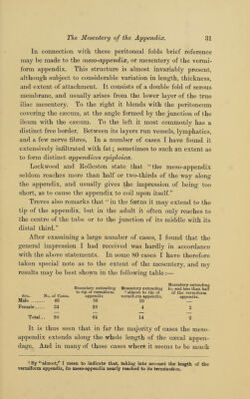
![:]2 The Meso-apjoendix. shortened, careful examination will often demonstrate a thin peritoneal fold extending up to its termination. Sometimes the mesentery appears to extend even beyond the termination of the appendix, and in some instances this terminal portion may present one or more large rounded collections of adipose tissue, projecting well beyond the end of the vermiform appendix. Fig. 10.—Variations in extent and attachment of the meso-appendix to a retro- cecal vermiform appendix. (Treves.) A.—Caecum turned upwards and showing the vermiform appendix in a retro-caecal position, and having a well-marked meso-appendix. B.—Retro-caecal appendix with its meso-appendix almost vertical, and having its attach- ment to the caecum only. C.—Caecum turned upwards and snowing the vermiform appendix in a retro-caecal position, and with diminished extent of mesentery. D.— Retro-csecal appendix, almost wholly adherent to the caecum, with greatly reduced meso-appendix. From a specimen taken from the body of a man, aged 33. The general appearance of the meso-appendix is illustrated in Figs. 6, 7, 8 and 10,' In Fig. 10 certain of the variations in the extent and attach- ment of the mesentery of the appendix, are shown.](https://iiif.wellcomecollection.org/image/b21061749_0048.jp2/full/800%2C/0/default.jpg)
 No text description is available for this image
 No text description is available for this image
 No text description is available for this image